Anti-ICOS Ligand/ICOSL antibody (ab138354)
Key features and details
- Rabbit polyclonal to ICOS Ligand/ICOSL
- Suitable for: WB, IHC-P
- Reacts with: Mouse
- Isotype: IgG
Overview
-
Product name
Anti-ICOS Ligand/ICOSL antibody
See all ICOS Ligand/ICOSL primary antibodies -
Description
Rabbit polyclonal to ICOS Ligand/ICOSL -
Host species
Rabbit -
Tested Applications & Species
See all applications and species dataApplication Species IHC-P MouseWB Mouse -
Immunogen
Synthetic peptide within Mouse ICOS Ligand/ICOSL aa 300 to the C-terminus conjugated to keyhole limpet haemocyanin. The exact sequence is proprietary.
Database link: Q9JHJ8 -
Positive control
- This antibody gave a positive signal in Mouse Heart and Kidney tissue lysates. This antibody gave a positive result in IHC in the following FFPE tissue: Mouse normal spleen.
-
General notes
This product was previously labelled as ICOS Ligand
Properties
-
Form
Liquid -
Storage instructions
Shipped at 4°C. Store at +4°C short term (1-2 weeks). Upon delivery aliquot. Store at -20°C or -80°C. Avoid freeze / thaw cycle. -
Storage buffer
pH: 7.40
Preservative: 0.02% Sodium azide
Constituent: PBS
Batches of this product that have a concentration Concentration information loading...
Purity
Immunogen affinity purifiedClonality
PolyclonalIsotype
IgGResearch areas
Associated products
-
Compatible Secondaries
-
Isotype control
-
Recombinant Protein
Applications
The Abpromise guarantee
Our Abpromise guarantee covers the use of ab138354 in the following tested applications.
The application notes include recommended starting dilutions; optimal dilutions/concentrations should be determined by the end user.
GuaranteedTested applications are guaranteed to work and covered by our Abpromise guarantee.
PredictedPredicted to work for this combination of applications and species but not guaranteed.
IncompatibleDoes not work for this combination of applications and species.
Application Species IHC-P MouseWB MouseApplication Abreviews Notes WB Use a concentration of 1 µg/ml. Detects a band of approximately 42 kDa (predicted molecular weight: 33 kDa).IHC-P (1) Use a concentration of 5 µg/ml. Perform heat mediated antigen retrieval with citrate buffer pH 6 before commencing with IHC staining protocol.Notes WB
Use a concentration of 1 µg/ml. Detects a band of approximately 42 kDa (predicted molecular weight: 33 kDa).IHC-P
Use a concentration of 5 µg/ml. Perform heat mediated antigen retrieval with citrate buffer pH 6 before commencing with IHC staining protocol.Target
-
Function
Ligand for the T-cell-specific cell surface receptor ICOS. Acts as a costimulatory signal for T-cell proliferation and cytokine secretion; induces also B-cell proliferation and differentiation into plasma cells. Could play an important role in mediating local tissue responses to inflammatory conditions, as well as in modulating the secondary immune response by co-stimulating memory T-cell function. -
Tissue specificity
Isoform 1 is widely expressed (brain, heart, kidney, liver, lung, pancreas, placenta, skeletal muscle, bone marrow, colon, ovary, prostate, testis, lymph nodes, leukocytes, spleen, thymus and tonsil), while isoform 2 is detected only in lymph nodes, leukocytes and spleen. Expressed on activated monocytes and dendritic cells. -
Sequence similarities
Belongs to the immunoglobulin superfamily. BTN/MOG family.
Contains 1 Ig-like C2-type (immunoglobulin-like) domain.
Contains 1 Ig-like V-type (immunoglobulin-like) domain. -
Cellular localization
Membrane. - Information by UniProt
-
Database links
- Entrez Gene: 50723 Mouse
- SwissProt: Q9JHJ8 Mouse
- Unigene: 17819 Mouse
-
Alternative names
- B7 H2 antibody
- B7 homolog 2 antibody
- B7 homologue 2 antibody
see all
Images
-
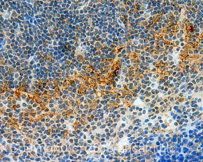
 Immunohistochemistry (Formalin/PFA-fixed paraffin-embedded sections) - Anti-ICOS Ligand/ICOSL antibody (ab138354)
IHC image of ICOS Ligand/ICOSL staining in Mouse normal spleen formalin fixed paraffin embedded tissue section, performed on a Leica Bond™ system using the standard protocol B. The section was pre-treated using heat mediated antigen retrieval with sodium citrate buffer (pH6, epitope retrieval solution 1) for 20 mins. The section was then incubated with ab138354, 5µg/ml, for 15 mins at room temperature. A Goat anti-Rabbit biotinylated secondary antibody was used to detect the primary, and visualized using an HRP conjugated ABC system. DAB was used as the chromogen. The section was then counterstained with haematoxylin and mounted with DPX.
For other IHC staining systems (automated and non-automated) customers should optimize variable parameters such as antigen retrieval conditions, primary antibody concentration and antibody incubation times.
-
All lanes : Anti-ICOS Ligand/ICOSL antibody (ab138354) at 1 µg/ml
Lane 1 : Heart (Mouse) Tissue Lysate
Lane 2 : Kidney (Mouse) Tissue Lysate
Lane 3 : Heart (Mouse) Tissue Lysate with Immunising peptide at 1 µg/ml
Lane 4 : Kidney (Mouse) Tissue Lysate with Immunising peptide at 1 µg/ml
Lysates/proteins at 25 µg per lane.
Secondary
All lanes : Goat Anti-Rabbit IgG H&L (HRP) (ab97051) at 1/10000 dilution
Developed using the ECL technique.
Performed under reducing conditions.
Predicted band size: 33 kDa
Observed band size: 42 kDa why is the actual band size different from the predicted?
Additional bands at: 28 kDa (possible non-specific binding)
Exposure time: 12 minutesThis blot was produced using a 10% Bis-tris gel under the MOPS buffer system. The gel was run at 200V for 50 minutes before being transferred onto a Nitrocellulose membrane at 30V for 70 minutes. The membrane was then blocked for an hour using 5% Bovine Serum Albumin before being incubated with ab138354 overnight at 4°C. Antibody binding was detected using an anti-rabbit antibody conjugated to HRP, and visualised using ECL development solution.
Protocols
References (4)
ab138354 has been referenced in 4 publications.
- Knox T et al. Selective HDAC6 inhibitors improve anti-PD-1 immune checkpoint blockade therapy by decreasing the anti-inflammatory phenotype of macrophages and down-regulation of immunosuppressive proteins in tumor cells. Sci Rep 9:6136 (2019). PubMed: 30992475
- Roth M et al. Pelargonium sidoides radix extract EPs 7630 reduces rhinovirus infection through modulation of viral binding proteins on human bronchial epithelial cells. PLoS One 14:e0210702 (2019). PubMed: 30707726
- Roth M et al. Broncho Vaxom (OM-85) modulates rhinovirus docking proteins on human airway epithelial cells via Erk1/2 mitogen activated protein kinase and cAMP. PLoS One 12:e0188010 (2017). PubMed: 29182620
- Lownik JC et al. ADAM10-Mediated ICOS Ligand Shedding on B Cells Is Necessary for Proper T Cell ICOS Regulation and T Follicular Helper Responses. J Immunol 199:2305-2315 (2017). PubMed: 28814605
Images
-
 Immunohistochemistry (Formalin/PFA-fixed paraffin-embedded sections) - Anti-ICOS Ligand/ICOSL antibody (ab138354)
IHC image of ICOS Ligand/ICOSL staining in Mouse normal spleen formalin fixed paraffin embedded tissue section, performed on a Leica Bond™ system using the standard protocol B. The section was pre-treated using heat mediated antigen retrieval with sodium citrate buffer (pH6, epitope retrieval solution 1) for 20 mins. The section was then incubated with ab138354, 5µg/ml, for 15 mins at room temperature. A Goat anti-Rabbit biotinylated secondary antibody was used to detect the primary, and visualized using an HRP conjugated ABC system. DAB was used as the chromogen. The section was then counterstained with haematoxylin and mounted with DPX.
For other IHC staining systems (automated and non-automated) customers should optimize variable parameters such as antigen retrieval conditions, primary antibody concentration and antibody incubation times.
-
All lanes : Anti-ICOS Ligand/ICOSL antibody (ab138354) at 1 µg/ml
Lane 1 : Heart (Mouse) Tissue Lysate
Lane 2 : Kidney (Mouse) Tissue Lysate
Lane 3 : Heart (Mouse) Tissue Lysate with Immunising peptide at 1 µg/ml
Lane 4 : Kidney (Mouse) Tissue Lysate with Immunising peptide at 1 µg/ml
Lysates/proteins at 25 µg per lane.
Secondary
All lanes : Goat Anti-Rabbit IgG H&L (HRP) (ab97051) at 1/10000 dilution
Developed using the ECL technique.
Performed under reducing conditions.
Predicted band size: 33 kDa
Observed band size: 42 kDa why is the actual band size different from the predicted?
Additional bands at: 28 kDa (possible non-specific binding)
Exposure time: 12 minutesThis blot was produced using a 10% Bis-tris gel under the MOPS buffer system. The gel was run at 200V for 50 minutes before being transferred onto a Nitrocellulose membrane at 30V for 70 minutes. The membrane was then blocked for an hour using 5% Bovine Serum Albumin before being incubated with ab138354 overnight at 4°C. Antibody binding was detected using an anti-rabbit antibody conjugated to HRP, and visualised using ECL development solution.


