Anti-Iba1 antibody (ab108539)
")
Key features and details
- Rabbit polyclonal to Iba1
- Suitable for: WB, IHC-P
- Reacts with: Mouse, Rat, Human
- Isotype: IgG
Overview
-
Product name
Anti-Iba1 antibody
See all Iba1 primary antibodies -
Description
Rabbit polyclonal to Iba1 -
Host species
Rabbit -
Specificity
In WB, we recommend blocking in milk. Blocking with BSA gives high background. If looking for a monoclonal anti-Iba1 alternative we can recommend our RabMAb ab178846 -
Tested applications
Suitable for: WB, IHC-Pmore details -
Species reactivity
Reacts with: Mouse, Rat, Human -
Immunogen
Synthetic peptide corresponding to Human Iba1 (C terminal).
Database link: P55008 -
Positive control
- Iba1 (Human) full length recombinant protein; Rat brain lysate; Human fetal lymphocytes; spleen tissue lysate; THP1, C6, NR8383 whole cell lysate
-
General notes
Reproducibility is key to advancing scientific discovery and accelerating scientists’ next breakthrough.
Abcam is leading the way with our range of recombinant antibodies, knockout-validated antibodies and knockout cell lines, all of which support improved reproducibility.
We are also planning to innovate the way in which we present recommended applications and species on our product datasheets, so that only applications & species that have been tested in our own labs, our suppliers or by selected trusted collaborators are covered by our Abpromise™ guarantee.
In preparation for this, we have started to update the applications & species that this product is Abpromise guaranteed for.
We are also updating the applications & species that this product has been “predicted to work with,” however this information is not covered by our Abpromise guarantee.
Applications & species from publications and Abreviews that have not been tested in our own labs or in those of our suppliers are not covered by the Abpromise guarantee.
Please check that this product meets your needs before purchasing. If you have any questions, special requirements or concerns, please send us an inquiry and/or contact our Support team ahead of purchase. Recommended alternatives for this product can be found below, as well as customer reviews and Q&As.
Properties
-
Form
Lyophilized:Reconstitute with 200ul distilled sterile water. Please note that if you receive this product in liquid form it has already been reconstituted as described and no further reconstitution is necessary. -
Storage instructions
Shipped at 4°C. Upon delivery aliquot and store at -20°C. Avoid repeated freeze / thaw cycles. -
Storage buffer
pH: 7.20
Preservative: 0.02% Sodium azide
Constituents: PBS, 1% BSA -
 Concentration information loading...
 -
Purity
Immunogen affinity purified -
Clonality
Polyclonal -
Isotype
IgG -
Research areas
Images
-
Immunohistochemistry (Formalin/PFA-fixed paraffin-embedded sections) - Anti-Iba1 antibody (ab108539)
IHC image of Iba1 staining in normal mouse brain formalin fixed paraffin embedded tissue section, performed on a Leica Bond™ system using the standard protocol B. The section was pre-treated using heat mediated antigen retrieval with sodium citrate buffer (pH6, epitope retrieval solution 1) for 20 mins. The section was then incubated with ab108539, 1/1000 dilution, for 15 mins at room temperature and detected using an HRP conjugated ABC system. DAB was used as the chromogen. The section was then counterstained with haematoxylin and mounted with DPX.
For other IHC staining systems (automated and non-automated) customers should optimize variable parameters such as antigen retrieval conditions, primary antibody concentration and antibody incubation times.
-
 Immunohistochemistry (Formalin/PFA-fixed paraffin-embedded sections) - Anti-Iba1 antibody (ab108539)
IHC image of Iba1 staining in normal rat brain formalin fixed paraffin embedded tissue section, performed on a Leica Bond™ system using the standard protocol F. The section was pre-treated using heat mediated antigen retrieval with sodium citrate buffer (pH6, epitope retrieval solution 1) for 20 mins. The section was then incubated with ab108539, 1/500 dilution, for 15 mins at room temperature and detected using an HRP conjugated compact polymer system. DAB was used as the chromogen. The section was then counterstained with haematoxylin and mounted with DPX.
For other IHC staining systems (automated and non-automated) customers should optimize variable parameters such as antigen retrieval conditions, primary antibody concentration and antibody incubation times.
-
 Immunohistochemistry (Formalin/PFA-fixed paraffin-embedded sections) - Anti-Iba1 antibody (ab108539)
IHC image of Iba1 staining in normal human hippocampus formalin fixed paraffin embedded tissue section, performed on a Leica Bond™ system using the standard protocol F. The section was pre-treated using heat mediated antigen retrieval with sodium citrate buffer (pH6, epitope retrieval solution 1) for 20 mins. The section was then incubated with ab108539, 1/1000 dilution, for 15 mins at room temperature and detected using an HRP conjugated compact polymer system. DAB was used as the chromogen. The section was then counterstained with haematoxylin and mounted with DPX.
For other IHC staining systems (automated and non-automated) customers should optimize variable parameters such as antigen retrieval conditions, primary antibody concentration and antibody incubation times.
-
 Immunohistochemistry (Formalin/PFA-fixed paraffin-embedded sections) - Anti-Iba1 antibody (ab108539)ab108539, at 1/100 dilution, staining Iba1 in formalin-fixed, paraffin-embedded Human fetal lymphocytes by Immunohistochemistry.
 -
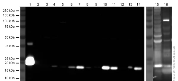 Western blot - Anti-Iba1 antibody (ab108539)All lanes : Anti-Iba1 antibody (ab108539) at 1/1000 dilution
Lane 1 : Human Iba1 recombinant protein at 0.1 µg
Lane 2 : HEK293 whole cell lysate at 20 µg
Lane 3 : A431 whole cell lysate at 20 µg
Lane 4 : NIH3T3 whole cell lysate at 30 µg
Lane 5 : Human spleen tissue lysate at 20 µg
Lane 6 : Mouse spleen tissue lysate at 30 µg
Lane 7 : Rat spleen tissue lysate at 30 µg
Lane 8 : U937 whole cell lysate at 30 µg
Lane 9 : MOLT4 whole cell lysate at 20 µg
Lane 10 : THP1 whole cell lysate at 30 µg
Lane 11 : THP1 whole cell lysate, PMA treated at 30 µg
Lane 12 : Raw 264.7 whole cell lysate at 30 µg
Lane 13 : C6 whole cell lysate at 30 µg
Lane 14 : NR8383 whole cell lysate at 30 µg
Lane 15 : NIH3T3 whole cell lysate - BLOCKED IN 5% BSA at 30 µg
Lane 16 : Human spleen tissue lysate - BLOCKED IN 5% BSA at 20 µg
Developed using the ECL technique.
Performed under reducing conditions.
Predicted band size: 17 kDa
Exposure time: 3 minutesLanes 1-14: Blocked in 3% milk for 1 hour (RT). Lanes 15-16: Blocked in 5% BSA for 1 hour (RT).
Abcam recommends blocking in milk for cleaner blots with reduced background, in comparison to BSA.
This blot was produced using a 4-12% Bis-Tris gel under the MOPS buffer system. The gel was run at 200V for 60 minutes before being transferred onto a nitrocellulose membrane at 30V for 70 minutes. The membrane was then blocked for an hour before being incubated with ab108539 (anti-Iba1 antibody; 1/1000) for 18 hours at 4°C. Antibody binding was detected using HRP-labelled anti-Rabbit IgG for 1 hour at room temperature and visualised using ECL development solution ab133406.
-
 Western blot - Anti-Iba1 antibody (ab108539)Anti-Iba1 antibody (ab108539) at 1/500 dilution + Rat brain lysate
Predicted band size: 17 kDa


