Anti-Galectin 3 antibody (ab31707)
Key features and details
- Rabbit polyclonal to Galectin 3
- Suitable for: ICC/IF, WB
- Knockout validated
- Reacts with: Human
- Isotype: IgG
Overview
-
Product name
Anti-Galectin 3 antibody
See all Galectin 3 primary antibodies -
Description
Rabbit polyclonal to Galectin 3 -
Host species
Rabbit -
Tested Applications & Species
See all applications and species dataApplication Species ICC/IF HumanWB Human -
Immunogen
Synthetic peptide corresponding to Human Galectin 3 aa 200 to the C-terminus (C terminal) conjugated to keyhole limpet haemocyanin.
(Peptide available asab31706) -
Positive control
- Recombinant human Galectin 3 protein (ab50236) can be used as a positive control in WB. HeLa (Human epithelial carcinoma cell line) Whole Cell and Nuclear Lysates.
-
General notes
The Life Science industry has been in the grips of a reproducibility crisis for a number of years. Abcam is leading the way in addressing the problem with our range of recombinant monoclonal antibodies and knockout edited cell lines for gold-standard validation.
One factor contributing to the crisis is the use of antibodies that are not suitable. This can lead to misleading results and the use of incorrect data informing project assumptions and direction. To help address this challenge, we have introduced an application and species grid on our primary antibody datasheets to make it easy to simplify identification of the right antibody for your needs.
Learn more here.
Properties
-
Form
Liquid -
Storage instructions
Shipped at 4°C. Store at +4°C short term (1-2 weeks). Upon delivery aliquot. Store at -20°C or -80°C. Avoid freeze / thaw cycle. -
Storage buffer
pH: 7.40
Preservative: 0.02% Sodium azide
Constituent: PBS
Batches of this product that have a concentration Concentration information loading...
Purity
Immunogen affinity purifiedClonality
PolyclonalIsotype
IgGResearch areas
Associated products
-
Compatible Secondaries
-
Isotype control
-
Positive Controls
-
Recombinant Protein
Applications
The Abpromise guarantee
Our Abpromise guarantee covers the use of ab31707 in the following tested applications.
The application notes include recommended starting dilutions; optimal dilutions/concentrations should be determined by the end user.
GuaranteedTested applications are guaranteed to work and covered by our Abpromise guarantee.
PredictedPredicted to work for this combination of applications and species but not guaranteed.
IncompatibleDoes not work for this combination of applications and species.
Application Species ICC/IF HumanWB HumanAll applications DogPigApplication Abreviews Notes ICC/IF (1) Use a concentration of 1 µg/ml.WB (2) Use a concentration of 1 µg/ml. Detects a band of approximately 30 kDa (predicted molecular weight: 26 kDa).Notes ICC/IF
Use a concentration of 1 µg/ml.WB
Use a concentration of 1 µg/ml. Detects a band of approximately 30 kDa (predicted molecular weight: 26 kDa).Target
-
Function
Galactose-specific lectin which binds IgE. May mediate with the alpha-3, beta-1 integrin the stimulation by CSPG4 of endothelial cells migration. Together with DMBT1, required for terminal differentiation of columnar epithelial cells during early embryogenesis. -
Tissue specificity
A major expression is found in the colonic epithelium. It is also abundant in the activated macrophages. -
Sequence similarities
Contains 1 galectin domain. -
Cellular localization
Nucleus. Cytoplasmic in adenomas and carcinomas. May be secreted by a non-classical secretory pathway and associate with the cell surface. - Information by UniProt
-
Database links
- Entrez Gene: 3958 Human
- Omim: 153619 Human
- SwissProt: P38486 Dog
- SwissProt: P17931 Human
- Unigene: 531081 Human
-
Alternative names
- 35 kDa lectin antibody
- Carbohydrate binding protein 35 antibody
- Carbohydrate-binding protein 35 antibody
see all
Images
-
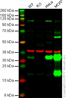
Lane 1: Wild-type HAP1 whole cell lysate (20 µg)
Lane 2: Galectin 3 knockout HAP1 whole cell lysate (20 µg)
Lane 3: HeLa whole cell lysate (20 µg)
Lane 4: MCF7 whole cell lysate (20 µg)Lanes 1 - 4: Merged signal (red and green). Green - ab31707 observed at 32 kDa. Red - loading control, ab8245, observed at 37 kDa.
ab31707 was shown to specifically recognize Galectin 3 in wild-type HAP1 cells along with additional cross-reactive bands. No band was observed when Galectin 3 knockout samples were examined. Wild-type and Galectin 3 knockout samples were subjected to SDS-PAGE. Ab31707 and ab8245 (Mouse anti GAPDH loading control) were incubated overnight at 4°C at 1 ug/ml and 1/10000 dilution respectively. Blots were developed with Goat anti-Rabbit IgG H&L (IRDye® 800CW) preabsorbed ab216773 and Goat anti-Mouse IgG H&L (IRDye® 680RD) preabsorbed ab216776 secondary antibodies at 1/20000 dilution for 1 hour at room temperature before imaging.
-
ICC/IF image of ab31707 stained human HeLa cells. The cells were PFA fixed (10 min), permabilised in TBS-T (20 min) and incubated with the antibody (ab31707, 1µg/ml) for 1h at room temperature. 1%BSA / 10% normal goat serum / 0.3M glycine was used to quench autofluorescence and block non-specific protein-protein interactions. The secondary antibody (green) was Alexa Fluor® 488 goat anti-rabbit IgG (H+L) used at a 1/1000 dilution for 1h. Alexa Fluor® 594 WGA was used to label plasma membranes (red). DAPI was used to stain the cell nuclei (blue).
-
All lanes : Anti-Galectin 3 antibody (ab31707) at 1 µg/ml
Lane 1 : HeLa (Human epithelial carcinoma cell line) Whole Cell Lysate
Lane 2 : HeLa (Human epithelial carcinoma cell line) Nuclear Lysate
Lysates/proteins at 20 µg per lane.
Secondary
All lanes : IRDye 680 Conjugated Goat Anti Rabbit IgG (H&L) at 1/15000 dilution
Performed under reducing conditions.
Predicted band size: 26 kDa
Observed band size: 30 kDa why is the actual band size different from the predicted?
We also see a very faint band at 52 kDa in the HeLa Whole cell lysate, we are uncertain as to the identity of this band.
Protocols
Datasheets and documents
-
SDS download
-
Datasheet download
References (9)
ab31707 has been referenced in 9 publications.
- Koerver L et al. The ubiquitin-conjugating enzyme UBE2QL1 coordinates lysophagy in response to endolysosomal damage. EMBO Rep 20:e48014 (2019). PubMed: 31432621
- Coppin L et al. Galectin-3 is a non-classic RNA binding protein that stabilizes the mucin MUC4 mRNA in the cytoplasm of cancer cells. Sci Rep 7:43927 (2017). PubMed: 28262838
- Zheng J et al. Galectin-3 induced by hypoxia promotes cell migration in thyroid cancer cells. Oncotarget 8:101475-101488 (2017). PubMed: 29254179
- Okawa S et al. Proteome and Secretome Characterization of Glioblastoma-Derived Neural Stem Cells. Stem Cells 35:967-980 (2017). PubMed: 27870168
- Truchan HK et al. Type II Secretion Substrates of Legionella pneumophila Translocate Out of the Pathogen-Occupied Vacuole via a Semipermeable Membrane. MBio 8:N/A (2017). ICC/IF . PubMed: 28634242
- Ma Z et al. Galectin-3 Inhibition Is Associated with Neuropathic Pain Attenuation after Peripheral Nerve Injury. PLoS One 11:e0148792 (2016). WB . PubMed: 26872020
- DE Oliveira JT et al. Differential expression of galectin-1 and galectin-3 in canine non-malignant and malignant mammary tissues and in progression to metastases in mammary tumors. Anticancer Res 34:2211-21 (2014). PubMed: 24778023
- Petropolis DB et al. A new human 3D-liver model unravels the role of galectins in liver infection by the parasite Entamoeba histolytica. PLoS Pathog 10:e1004381 (2014). ICC/IF ; Human . PubMed: 25211477
- Cui GQ et al. Proteomic analysis of meningiomas. Acta Neurol Belg 114:187-94 (2014). WB ; Human . PubMed: 24085542
Images
-
Lane 1: Wild-type HAP1 whole cell lysate (20 µg)
Lane 2: Galectin 3 knockout HAP1 whole cell lysate (20 µg)
Lane 3: HeLa whole cell lysate (20 µg)
Lane 4: MCF7 whole cell lysate (20 µg)Lanes 1 - 4: Merged signal (red and green). Green - ab31707 observed at 32 kDa. Red - loading control, ab8245, observed at 37 kDa.
ab31707 was shown to specifically recognize Galectin 3 in wild-type HAP1 cells along with additional cross-reactive bands. No band was observed when Galectin 3 knockout samples were examined. Wild-type and Galectin 3 knockout samples were subjected to SDS-PAGE. Ab31707 and ab8245 (Mouse anti GAPDH loading control) were incubated overnight at 4°C at 1 ug/ml and 1/10000 dilution respectively. Blots were developed with Goat anti-Rabbit IgG H&L (IRDye® 800CW) preabsorbed ab216773 and Goat anti-Mouse IgG H&L (IRDye® 680RD) preabsorbed ab216776 secondary antibodies at 1/20000 dilution for 1 hour at room temperature before imaging.
-
ICC/IF image of ab31707 stained human HeLa cells. The cells were PFA fixed (10 min), permabilised in TBS-T (20 min) and incubated with the antibody (ab31707, 1µg/ml) for 1h at room temperature. 1%BSA / 10% normal goat serum / 0.3M glycine was used to quench autofluorescence and block non-specific protein-protein interactions. The secondary antibody (green) was Alexa Fluor® 488 goat anti-rabbit IgG (H+L) used at a 1/1000 dilution for 1h. Alexa Fluor® 594 WGA was used to label plasma membranes (red). DAPI was used to stain the cell nuclei (blue).
-
All lanes : Anti-Galectin 3 antibody (ab31707) at 1 µg/ml
Lane 1 : HeLa (Human epithelial carcinoma cell line) Whole Cell Lysate
Lane 2 : HeLa (Human epithelial carcinoma cell line) Nuclear Lysate
Lysates/proteins at 20 µg per lane.
Secondary
All lanes : IRDye 680 Conjugated Goat Anti Rabbit IgG (H&L) at 1/15000 dilution
Performed under reducing conditions.
Predicted band size: 26 kDa
Observed band size: 30 kDa why is the actual band size different from the predicted?
We also see a very faint band at 52 kDa in the HeLa Whole cell lysate, we are uncertain as to the identity of this band.


