Anti-Galectin 1 antibody (ab25138)
")
Key features and details
- Rabbit polyclonal to Galectin 1
- Suitable for: IHC-P, ICC/IF, WB
- Reacts with: Human
- Isotype: IgG
Overview
-
Product name
Anti-Galectin 1 antibody
See all Galectin 1 primary antibodies -
Description
Rabbit polyclonal to Galectin 1 -
Host species
Rabbit -
Tested applications
Suitable for: IHC-P, ICC/IF, WBmore details -
Species reactivity
Reacts with: Human -
Immunogen
Recombinant full length protein (Human).
Properties
-
Form
Liquid -
Storage instructions
Shipped at 4°C. Store at +4°C short term (1-2 weeks). Upon delivery aliquot. Store at -20°C or -80°C. Avoid freeze / thaw cycle. -
Storage buffer
Constituent: 1.34% PBS -
 Concentration information loading...
 -
Purity
Immunogen affinity purified -
Clonality
Polyclonal -
Isotype
IgG -
Research areas
Images
-
Western blot - Anti-Galectin 1 antibody (ab25138)Anti-Galectin 1 antibody (ab25138) at 1 µg/ml + Human placenta tissue lysate - total protein (ab29745) at 10 µg
Secondary
Goat Anti-Rabbit IgG H&L (HRP) preadsorbed (ab97080) at 1/5000 dilution
Developed using the ECL technique.
Performed under reducing conditions.
Predicted band size: 14 kDa
Observed band size: 14 kDa
Additional bands at: 32 kDa, 45 kDa, 73 kDa. We are unsure as to the identity of these extra bands.
Exposure time: 30 seconds
-
 Immunocytochemistry/ Immunofluorescence - Anti-Galectin 1 antibody (ab25138)ICC/IF image of ab25138 stained Hek293 cells. The cells were 4% PFA fixed (10 min) and then incubated in 1%BSA / 10% normal goat serum / 0.3M glycine in 0.1% PBS-Tween for 1h to permeabilise the cells and block non-specific protein-protein interactions. The cells were then incubated with the antibody (ab25138, 1µg/ml) overnight at +4°C. The secondary antibody (green) was Alexa Fluor® 488 goat anti-rabbit IgG (H+L) used at a 1/1000 dilution for 1h. Alexa Fluor® 594 WGA was used to label plasma membranes (red) at a 1/200 dilution for 1h. DAPI was used to stain the cell nuclei (blue) at a concentration of 1.43µM.
-
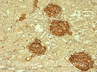 Immunohistochemistry (Formalin/PFA-fixed paraffin-embedded sections) - Anti-Galectin 1 antibody (ab25138)IHC image of ab25138 staining in human normal kidney formalin fixed paraffin embedded tissue section, performed on a Leica BondTM system using the standard protocol F. The section was pre-treated using heat mediated antigen retrieval with sodium citrate buffer (pH6, epitope retrieval solution 1) for 20 mins. The section was then incubated with ab25138, 1µg/ml, for 15 mins at room temperature and detected using an HRP conjugated compact polymer system. DAB was used as the chromogen. The section was then counterstained with haematoxylin and mounted with DPX.
For other IHC staining systems (automated and non-automated) customers should optimize variable parameters such as antigen retrieval conditions, primary antibody concentration and antibody incubation times.
-
 Immunohistochemistry (Formalin/PFA-fixed paraffin-embedded sections) - Anti-Galectin 1 antibody (ab25138) Punt S et al.; PLoS One. 2015; 10(6): e0129119., Fig 3, doi: 10.1371/journal.pone.0129119 Reproduced under the Creative Commons license http://creativecommons.org/licenses/by/4.0/
ab25138 staining Galectin-1 in Human cervical cancer tissue sections by Immunohistochemistry (IHC-P - paraformaldehyde-fixed, paraffin-embedded sections). Samples were incubated with primary antibody (1/1000 in 1% BSA in PBS) overnight at room temperature. An Alexa Fluor A647-conjugated Donkey anti-rabbit polyclonal (1/200) was used as the secondary antibody.
Representative images from double stainings of Galectin-1 and CD163. Arrows indicate examples of double positive cells.
-
 Immunohistochemistry (Formalin/PFA-fixed paraffin-embedded sections) - Anti-Galectin 1 antibody (ab25138) Punt S et al.; PLoS One. 2015; 10(6): e0129119., Fig 3, doi: 10.1371/journal.pone.0129119
ab25138 staining Galectin-1 in Human cervical cancer tissue sections by Immunohistochemistry (IHC-P - paraformaldehyde-fixed, paraffin-embedded sections). Samples were incubated with primary antibody (1/1000 in 1% BSA in PBS) overnight at room temperature. An Alexa Fluor A647-conjugated Donkey anti-rabbit polyclonal (1/200) was used as the secondary antibody.
Representative images from double stainings of Galectin-1 and CD3.


