Anti-GABA B Receptor 1 antibody (ab55051)
")
Key features and details
- Mouse monoclonal to GABA B Receptor 1
- Suitable for: WB, Flow Cyt, ICC/IF
- Reacts with: Human
- Isotype: IgG2a
Overview
-
Product name
Anti-GABA B Receptor 1 antibody
See all GABA B Receptor 1 primary antibodies -
Description
Mouse monoclonal to GABA B Receptor 1 -
Host species
Mouse -
Tested Applications & Species
See all applications and species dataApplication Species Flow Cyt HumanICC/IF HumanWB Human
-
Immunogen
Recombinant fragment: AVYIGALFPM SGGWPGGQAC QPAVEMALED VNSRRDILPD YELKLIHHDS KCDPGQATKY LYELLYNDPI KIILMPGCSS VSTLVAEAAR MWNLIVLSYG , corresponding to amino acids 52-151 of Human GABA B Receptor 1
-
General notes
This product was changed from ascites to tissue culture supernatant on 29th May 2019. Please note that the dilutions may need to be adjusted accordingly. If you have any questions, please do not hesitate to contact our scientific support team.
The Life Science industry has been in the grips of a reproducibility crisis for a number of years. Abcam is leading the way in addressing the problem with our range of recombinant monoclonal antibodies and knockout edited cell lines for gold-standard validation.
One factor contributing to the crisis is the use of antibodies that are not suitable. This can lead to misleading results and the use of incorrect data informing project assumptions and direction. To help address this challenge, we have introduced an application and species grid on our primary antibody datasheets to make it easy to simplify identification of the right antibody for your needs.
Learn more here.


Properties
-
Form
Liquid -
Storage instructions
Shipped at 4°C. Upon delivery aliquot and store at -20°C or -80°C. Avoid repeated freeze / thaw cycles. -
Storage buffer
pH: 7.4
Constituents: 8% Sodium chloride, 0.6% Dibasic monohydrogen sodium phosphate, 0.2% Monobasic dihydrogen potassium phosphate, 0.2% Potassium chloride, 91% Water -
 Concentration information loading...
 -
Purity
Tissue culture supernatant -
Clonality
Monoclonal -
Isotype
IgG2a -
Light chain type
kappa -
Research areas
Images
-
Immunocytochemistry/ Immunofluorescence - Anti-GABA B Receptor 1 antibody (ab55051)
ICC/IF image of ab55051 stained SHSY5Y cells. The cells were 4% formaldehyde fixed (10 min) and then incubated in 1%BSA / 10% normal goat serum / 0.3M glycine in 0.1% PBS-Tween for 1h to permeabilise the cells and block non-specific protein-protein interactions. The cells were then incubated with the antibody (ab55051, 5µg/ml) overnight at +4°C. The secondary antibody (green) was Alexa Fluor® 488 goat anti-mouse IgG (H+L) used at a 1/1000 dilution for 1h. Alexa Fluor® 594 WGA was used to label plasma membranes (red) at a 1/200 dilution for 1h. DAPI was used to stain the cell nuclei (blue) at a concentration of 1.43µM.
This image was generated using the ascites version of the product.
-
 Immunocytochemistry/ Immunofluorescence - Anti-GABA B Receptor 1 antibody (ab55051)
ab55051 staining GABA B receptor 1 in SK-N-SH cells treated with L-Glutamate (ab120049), by ICC/IF. Internalization of GABA B receptor 1 correlates with increased concentration of L-Glutamate, as described in literature.
The cells were incubated at 37°C for 30 minutes in media containing different concentrations of ab120049 (L-Glutamate) in DMSO, fixed with 4% formaldehyde for 10 minutes at room temperature and blocked with PBS containing 10% goat serum, 0.3 M glycine, 1% BSA and 0.1% tween for 2h at room temperature. Staining of the treated cells with ab55051 (1 µg/ml) was performed overnight at 4°C in PBS containing 1% BSA and 0.1% tween. A DyLight 488 goat anti-mouse polyclonal antibody (ab96879) at 1/250 dilution was used as the secondary antibody. Nuclei were counterstained with DAPI and are shown in blue.This image was generated using the ascites version of the product.
-
 Western blot - Anti-GABA B Receptor 1 antibody (ab55051)
GABA B Receptor 1 antibody (ab55051) at 1ug/lane + IMR-32 cell lysate at 25ug/lane.
This image was generated using the ascites version of the product.
-
 Flow Cytometry - Anti-GABA B Receptor 1 antibody (ab55051)
Overlay histogram showing SH-SY5Y cells stained with ab55051 (red line). The cells were fixed with 4% paraformaldehyde (10 min) and then permeabilized with 0.1% PBS-Tween for 20 min. The cells were then incubated in 1x PBS / 10% normal goat serum / 0.3M glycine to block non-specific protein-protein interactions followed by the antibody (ab55051, 0.5µg/1x106 cells) for 30 min at 22ºC. The secondary antibody used was DyLight® 488 goat anti-mouse IgG (H+L) (ab96879) at 1/500 dilution for 30 min at 22ºC. Isotype control antibody (black line) was mouse IgG2a [ICIGG2A] (ab91361, 1µg/1x106 cells) used under the same conditions. Acquisition of >5,000 events was performed. This antibody gave a positive signal in SH-SY5Y cells fixed with 80% methanol (5 min)/permeabilized with 0.1% PBS-Tween for 20 min used under the same conditions.
This image was generated using the ascites version of the product.
-
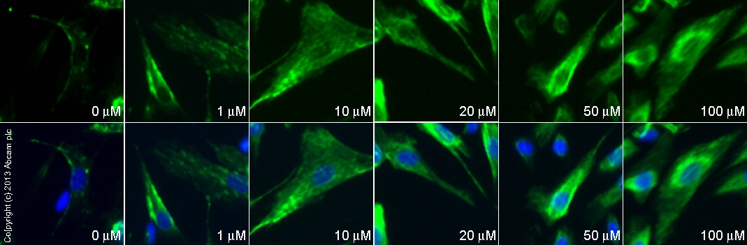
 Immunocytochemistry/ Immunofluorescence - Anti-GABA B Receptor 1 antibody (ab55051)
ab55051 staining GABA B receptor 1 in SK-N-SH cells treated with NMDA (ab120052), by ICC/IF. Internalization of GABA B receptor 1 correlates with increased concentration of NMDA, as described in literature.
The cells were incubated at 37°C for 30 minutes in media containing different concentrations of ab120052 (NMDA) in DMSO, fixed with 4% formaldehyde for 10 minutes at room temperature and blocked with PBS containing 10% goat serum, 0.3 M glycine, 1% BSA and 0.1% tween for 2h at room temperature. Staining of the treated cells with ab55051 (1 µg/ml) was performed overnight at 4°C in PBS containing 1% BSA and 0.1% tween. A DyLight 488 goat anti-mouse polyclonal antibody (ab96879) at 1/250 dilution was used as the secondary antibody. Nuclei were counterstained with DAPI and are shown in blue.This image was generated using the ascites version of the product.


