Anti-E2F1 (phospho T433) antibody (ab55325)
 antibody (ab55325)")
Key features and details
- Rabbit polyclonal to E2F1 (phospho T433)
- Suitable for: WB, IHC-P
- Reacts with: Human
- Isotype: IgG
Overview
-
Product name
Anti-E2F1 (phospho T433) antibody
See all E2F1 primary antibodies -
Description
Rabbit polyclonal to E2F1 (phospho T433) -
Host species
Rabbit -
Specificity
This antibody detects endogenous levels of E2F1 only when phosphorylated at threonine 433. -
Tested Applications & Species
See all applications and species dataApplication Species IHC-P HumanWB Human
-
Immunogen
Synthetic phosphopeptide derived from human E2F1 around the phosphorylation site of threonine 433 (D-L-TP-P-L).
-
Positive control
- HeLa cell extract.
Properties
-
Form
Liquid -
Storage instructions
Shipped at 4°C. Store at -20°C. Stable for 12 months at -20°C. -
Storage buffer
pH: 7.40
Preservative: 0.02% Sodium azide
Constituents: 0.87% Sodium chloride, 50% Glycerol (glycerin, glycerine), PBS -
 Concentration information loading...
 -
Purity
Immunogen affinity purified -
Purification notes
The antibody against non-phosphopeptide was removed by chromatography using non-phosphopeptide corresponding to the phosphorylation site. -
Clonality
Polyclonal -
Isotype
IgG -
Research areas
Images
-
Western blot - Anti-E2F1 (phospho T433) antibody (ab55325)All lanes : Anti-E2F1 (phospho T433) antibody (ab55325) at 1/500 dilution
Lane 1 : HeLa cell extract treated with Etoposide (at 25µM for 24 hrs).
Lane 2 : HeLa cell extract treated with Etoposide (at 25µM for 24 hrs), and with the immunising phosphopeptide.
Predicted band size: 47 kDa
Observed band size: 47 kDa
-
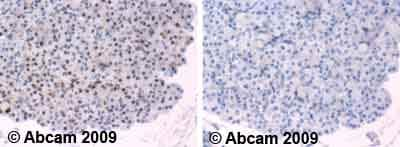 Immunohistochemistry (Formalin/PFA-fixed paraffin-embedded sections) - Anti-E2F1 (phospho T433) antibody (ab55325)Ab55325 staining human normal pancreas. Staining is localised to nuclear compartment.
Left panel: with primary antibody at 4 ug/ml. Right panel: isotype control.
Sections were stained using an automated system DAKO Autostainer Plus , at room temperature. Sections were rehydrated and antigen retrieved with the Dako 3-in-1 antigen retrieval buffer citrate pH6 in a DAKO PT Link. Slides were peroxidase blocked in 3% H2O2 in methanol for 10 minutes. They were then blocked with Dako Protein block for 10 minutes (containing casein 0.25% in PBS) then incubated with primary antibody for 20 minutes and detected with Dako Envision Flex amplification kit for 30 minutes. Colorimetric detection was completed with diaminobenzidine for 5 minutes. Slides were counterstained with Haematoxylin and coverslipped under DePeX. Please note that for manual staining we recommend to optimize the primary antibody concentration and incubation time (overnight incubation), and amplification may be required.

