Anti-Dcp2/TDT antibody (ab28658)
Key features and details
- Rabbit polyclonal to Dcp2/TDT
- Suitable for: ICC/IF, WB, IP
- Reacts with: Human
- Isotype: IgG
Overview
-
Product name
Anti-Dcp2/TDT antibody
See all Dcp2/TDT primary antibodies -
Description
Rabbit polyclonal to Dcp2/TDT -
Host species
Rabbit -
Tested applications
Suitable for: ICC/IF, WB, IPmore details -
Species reactivity
Reacts with: Human
Predicted to work with: Mouse
-
Immunogen
Synthetic peptide corresponding to Human Dcp2/TDT aa 300-400 conjugated to keyhole limpet haemocyanin.
(Peptide available asab30458) -
General notes
This product was previously labelled as Dcp2
Properties
-
Form
Liquid -
Storage instructions
Shipped at 4°C. Store at +4°C short term (1-2 weeks). Upon delivery aliquot. Store at -20°C or -80°C. Avoid freeze / thaw cycle. -
Storage buffer
pH: 7.40
Preservative: 0.02% Sodium azide
Constituent: PBS
Batches of this product that have a concentration Concentration information loading...
Purity
Immunogen affinity purifiedClonality
PolyclonalIsotype
IgGResearch areas
Associated products
-
Compatible Secondaries
-
Isotype control
-
Recombinant Protein
Applications
Our Abpromise guarantee covers the use of ab28658 in the following tested applications.
The application notes include recommended starting dilutions; optimal dilutions/concentrations should be determined by the end user.
Application Abreviews Notes ICC/IF Use a concentration of 1 µg/ml. WB Use a concentration of 1 µg/ml. Detects a band of approximately 52 kDa (predicted molecular weight: 48.4 kDa). IP Use at an assay dependent concentration. Target
-
Function
Necessary for the degradation of mRNAs, both in normal mRNA turnover and in nonsense-mediated mRNA decay. Plays a role in replication-dependent histone mRNA degradation. Removes the 7-methyl guanine cap structure from mRNA molecules, yielding a 5'-phosphorylated mRNA fragment and 7m-GDP. Has higher activity towards mRNAs that lack a poly(A) tail. Has no activity towards a cap structure lacking a RNA moiety. -
Sequence similarities
Belongs to the Nudix hydrolase family. DCP2 subfamily.
Contains 1 nudix hydrolase domain. -
Post-translational
modificationsPhosphorylated upon DNA damage, probably by ATM or ATR. -
Cellular localization
Cytoplasm > P-body. Nucleus. Predominantly cytoplasmic, in processing bodies (PB). A minor amount is nuclear. - Information by UniProt
-
Database links
- Entrez Gene: 167227 Human
- Entrez Gene: 70640 Mouse
- Omim: 609844 Human
- SwissProt: Q8IU60 Human
- SwissProt: Q9CYC6 Mouse
- Unigene: 443875 Human
- Unigene: 87629 Mouse
-
Alternative names
- DCP2 antibody
- DCP2 decapping enzyme homolog (S. cerevisiae) antibody
- DCP2, S. cerevisiae, homolog of antibody
see all
Images
-
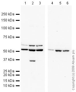
All lanes : Anti-Dcp2/TDT antibody (ab28658) at 1 µg/ml
Lane 1 : HeLa (Human epithelial carcinoma cell line) Whole Cell Lysate
Lane 2 : Jurkat (Human T cell lymphoblast-like cell line) Whole Cell Lysate
Lane 3 : HEK293 (Human embryonic kidney cell line) Whole Cell Lysate
Lane 4 : HeLa (Human epithelial carcinoma cell line) Whole Cell Lysate with Human Dcp2/TDT peptide (ab30458) at 1 µg/ml
Lane 5 : Jurkat (Human T cell lymphoblast-like cell line) Whole Cell Lysate with Human Dcp2/TDT peptide (ab30458) at 1 µg/ml
Lane 6 : HEK293 (Human embryonic kidney cell line) Whole Cell Lysate with Human Dcp2/TDT peptide (ab30458) at 1 µg/ml
Lysates/proteins at 10 µg per lane.
Secondary
All lanes : Goat polyclonal to Rabbit IgG - H&L - Pre-Adsorbed (HRP) at 1/3000 dilution
Performed under reducing conditions.
Predicted band size: 48.4 kDa
Observed band size: 52,58 kDa why is the actual band size different from the predicted?
Additional bands at: 35 kDa. We are unsure as to the identity of these extra bands.
Exposure time: 10 minutesDcp2/TDT contains a number of potential phosphorylation sites (Swiss Prot data) which may explain its migration at a higher molecular weight than predicted.
-
ICC/IF image of ab28658 stained human HeLa cells. The cells were PFA fixed (10 min), permabilised in TBS-T (20 min) and incubated with the antibody (ab28658, 1µg/ml) for 1h at room temperature. 1%BSA / 10% normal goat serum / 0.3M glycine was used to quench autofluorescence and block non-specific protein-protein interactions. The secondary antibody (green) was Alexa Fluor® 488 goat anti-rabbit IgG (H+L) used at a 1/1000 dilution for 1h. Alexa Fluor® 594 WGA was used to label plasma membranes (red). DAPI was used to stain the cell nuclei (blue).
-
Dcp2/TDT was immunoprecipitated using 0.5mg Jurkat whole cell extract, 5µg of Rabbit polyclonal to Dcp2/TDT and 50µl of protein G magnetic beads (+). No antibody was added to the control (-).
The antibody was incubated under agitation with Protein G beads for 10min, Jurkat whole cell extract lysate diluted in RIPA buffer was added to each sample and incubated for a further 10min under agitation.
Proteins were eluted by addition of 40µl SDS loading buffer and incubated for 10min at 70oC; 10µl of each sample was separated on a SDS PAGE gel, transferred to a nitrocellulose membrane, blocked with 5% BSA and probed with ab28658.
Secondary: Mouse monoclonal [SB62a] Secondary Antibody to Rabbit IgG light chain (HRP) (ab99697).
Band: 52,58kDa: Dcp2/TDT.
Protocols
References (4)
ab28658 has been referenced in 4 publications.
- Seto E et al. The Assembly of EDC4 and Dcp1a into Processing Bodies Is Critical for the Translational Regulation of IL-6. PLoS One 10:e0123223 (2015). WB . PubMed: 25970328
- Wang X et al. N6-methyladenosine-dependent regulation of messenger RNA stability. Nature 505:117-20 (2014). WB . PubMed: 24284625
- Chiang PY et al. Phosphorylation of mRNA decapping protein Dcp1a by the ERK signaling pathway during early differentiation of 3T3-L1 preadipocytes. PLoS One 8:e61697 (2013). WB ; Human . PubMed: 23637887
- Kim CW et al. Tristetraprolin controls the stability of cIAP2 mRNA through binding to the 3'UTR of cIAP2 mRNA. Biochem Biophys Res Commun 400:46-52 (2010). EMSA ; Human . PubMed: 20691152
Images
-
All lanes : Anti-Dcp2/TDT antibody (ab28658) at 1 µg/ml
Lane 1 : HeLa (Human epithelial carcinoma cell line) Whole Cell Lysate
Lane 2 : Jurkat (Human T cell lymphoblast-like cell line) Whole Cell Lysate
Lane 3 : HEK293 (Human embryonic kidney cell line) Whole Cell Lysate
Lane 4 : HeLa (Human epithelial carcinoma cell line) Whole Cell Lysate with Human Dcp2/TDT peptide (ab30458) at 1 µg/ml
Lane 5 : Jurkat (Human T cell lymphoblast-like cell line) Whole Cell Lysate with Human Dcp2/TDT peptide (ab30458) at 1 µg/ml
Lane 6 : HEK293 (Human embryonic kidney cell line) Whole Cell Lysate with Human Dcp2/TDT peptide (ab30458) at 1 µg/ml
Lysates/proteins at 10 µg per lane.
Secondary
All lanes : Goat polyclonal to Rabbit IgG - H&L - Pre-Adsorbed (HRP) at 1/3000 dilution
Performed under reducing conditions.
Predicted band size: 48.4 kDa
Observed band size: 52,58 kDa why is the actual band size different from the predicted?
Additional bands at: 35 kDa. We are unsure as to the identity of these extra bands.
Exposure time: 10 minutesDcp2/TDT contains a number of potential phosphorylation sites (Swiss Prot data) which may explain its migration at a higher molecular weight than predicted.
-
ICC/IF image of ab28658 stained human HeLa cells. The cells were PFA fixed (10 min), permabilised in TBS-T (20 min) and incubated with the antibody (ab28658, 1µg/ml) for 1h at room temperature. 1%BSA / 10% normal goat serum / 0.3M glycine was used to quench autofluorescence and block non-specific protein-protein interactions. The secondary antibody (green) was Alexa Fluor® 488 goat anti-rabbit IgG (H+L) used at a 1/1000 dilution for 1h. Alexa Fluor® 594 WGA was used to label plasma membranes (red). DAPI was used to stain the cell nuclei (blue).
-
Dcp2/TDT was immunoprecipitated using 0.5mg Jurkat whole cell extract, 5µg of Rabbit polyclonal to Dcp2/TDT and 50µl of protein G magnetic beads (+). No antibody was added to the control (-).
The antibody was incubated under agitation with Protein G beads for 10min, Jurkat whole cell extract lysate diluted in RIPA buffer was added to each sample and incubated for a further 10min under agitation.
Proteins were eluted by addition of 40µl SDS loading buffer and incubated for 10min at 70oC; 10µl of each sample was separated on a SDS PAGE gel, transferred to a nitrocellulose membrane, blocked with 5% BSA and probed with ab28658.
Secondary: Mouse monoclonal [SB62a] Secondary Antibody to Rabbit IgG light chain (HRP) (ab99697).
Band: 52,58kDa: Dcp2/TDT.


