Anti-Cytokeratin 19 antibody (ab53119)
")
Key features and details
- Rabbit polyclonal to Cytokeratin 19
- Suitable for: ICC/IF, WB, IHC-P
- Reacts with: Human
- Isotype: IgG
Overview
-
Product name
Anti-Cytokeratin 19 antibody
See all Cytokeratin 19 primary antibodies -
Description
Rabbit polyclonal to Cytokeratin 19 -
Host species
Rabbit -
Specificity
ab53119 detects endogenous levels of total Cytokeratin 19 protein. -
Tested applications
Suitable for: ICC/IF, WB, IHC-Pmore details -
Species reactivity
Reacts with: Human -
Immunogen
Synthetic peptide derived form human Cytokeratin 19.
Properties
-
Form
Liquid -
Storage instructions
Shipped at 4°C. Store at -20°C. Stable for 12 months at -20°C. -
Storage buffer
pH: 7.40
Preservative: 0.02% Sodium azide
Constituents: 50% Glycerol (glycerin, glycerine), 0.87% Sodium chloride, PBS
Without Mg+2 and Ca+2 -
 Concentration information loading...
 -
Purity
Immunogen affinity purified -
Clonality
Polyclonal -
Isotype
IgG -
Research areas
Images
-
Immunohistochemistry (Formalin/PFA-fixed paraffin-embedded sections) - Anti-Cytokeratin 19 antibody (ab53119)ab53119 at 1/50 dilution staining Cytokeratin 19 in human breast carcinoma by Immunohistochemistry, Paraffin embedded tissue, in the absence and presence of the immunising peptide.
-
 Western blot - Anti-Cytokeratin 19 antibody (ab53119)All lanes : Anti-Cytokeratin 19 antibody (ab53119) at 1/300 dilution
Lane 1 : LOVO cell extract
Lane 2 : LOVO cell extract with immunising peptide
Predicted band size: 44 kDa
Observed band size: 44 kDa
-
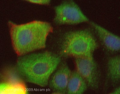 Immunocytochemistry/ Immunofluorescence - Anti-Cytokeratin 19 antibody (ab53119)ICC/IF image of ab53119 stained MCF7 cells. The cells were 4% PFA fixed (10 min) and then incubated in 1%BSA / 10% normal goat serum / 0.3M glycine in 0.1% PBS-Tween for 1h to permeabilise the cells and block non-specific protein-protein interactions. The cells were then incubated with the antibody (ab53119, 1µg/ml) overnight at +4°C. The secondary antibody (green) was Alexa Fluor® 488 goat anti-rabbit IgG (H+L) used at a 1/1000 dilution for 1h. Alexa Fluor® 594 WGA was used to label plasma membranes (red) at a 1/200 dilution for 1h. DAPI was used to stain the cell nuclei (blue) at a concentration of 1.43µM.


