Anti-Cleaved Caspase-3 antibody (ab2302)
Key features and details
- Rabbit polyclonal to Cleaved Caspase-3
- Suitable for: IHC-P, WB
- Reacts with: Human, Recombinant fragment
- Isotype: IgG
Overview
-
Product name
Anti-Cleaved Caspase-3 antibody
See all Cleaved Caspase-3 primary antibodies -
Description
Rabbit polyclonal to Cleaved Caspase-3 -
Host species
Rabbit -
Tested Applications & Species
See all applications and species dataApplication Species IHC-P HumanWB Human -
Immunogen
-
Positive control
- IHC-P: Human tonsil tissue. Human ulcerative colitis, colon tissue sections. WB: HeLa (treated with staurosporine) whole cell lysate. Camptothecin treated Jurkat whole cell lysate. Cleaved Caspase 3 recombinant protein.
-
General notes
Caspases are synthesized as inactive pro-enzymes that are processed to active form in cells undergoing apoptosis. Caspase 3 has been extensively studied and implicated to play an important role in apoptosis. Active caspase 3 proteolytically cleaves and activates other caspases, as well as relevant targets in the cells (e.g., PARP). This affinity purified antibody recognizing the active forms of caspase-3 provides a new tool for identifying apoptotic cell populations in both tissue sections and cultured cells.
Properties
-
Form
Liquid -
Storage instructions
Shipped at 4°C. Store at +4°C short term (1-2 weeks). Upon delivery aliquot. Store at -20°C or -80°C. Avoid freeze / thaw cycle. -
Storage buffer
Preservative: 0.03% Proclin 300
Constituents: PBS, 30% Glycerol (glycerin, glycerine), 0.5% BSA, 0.146% EDTA -
 Concentration information loading...
 -
Purity
Immunogen affinity purified -
Primary antibody notes
Caspases are synthesized as inactive pro-enzymes that are processed to active form in cells undergoing apoptosis. Caspase 3 has been extensively studied and implicated to play an important role in apoptosis. Active caspase 3 proteolytically cleaves and activates other caspases, as well as relevant targets in the cells (e.g., PARP). This affinity purified antibody recognizing the active forms of caspase-3 provides a new tool for identifying apoptotic cell populations in both tissue sections and cultured cells. -
Clonality
Polyclonal -
Isotype
IgG -
Research areas
Images
-
 Immunohistochemistry (Formalin/PFA-fixed paraffin-embedded sections) - Anti-Cleaved Caspase-3 antibody (ab2302)
IHC image of cleaved caspase-3 staining in human tonsil formalin fixed paraffin embedded tissue section*, performed on a Leica Bond™ system using the standard protocol F. The section was pre-treated using heat mediated antigen retrieval with sodium citrate buffer (pH6, epitope retrieval solution 1) for 20 mins. The section was then incubated with ab2302, 1µg/ml, for 15 mins at room temperature and detected using an HRP conjugated compact polymer system. DAB was used as the chromogen. The section was then counterstained with haematoxylin and mounted with DPX.
For other IHC staining systems (automated and non-automated) customers should optimize variable parameters such as antigen retrieval conditions, primary antibody concentration and antibody incubation times.
*Tissue obtained from the Human Research Tissue Bank, supported by the NIHR Cambridge Biomedical Research Centre
-
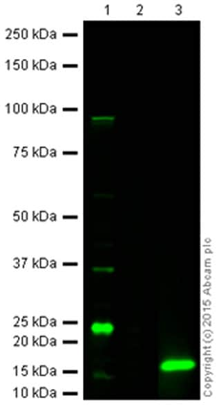
All lanes : Anti-Cleaved Caspase-3 antibody (ab2302) at 1 µg/ml
Lane 1 : HeLa (Human epithelial cell line from cervix adenocarcinoma) whole cell lysate (2 uM Staurosporine, 4Hr) at 20 µg
Lane 2 : HeLa whole cell lysate (untreated) at 20 µg
Lane 3 : Cleaved Caspase 3 (recombinant protein) at 0.1 µg
Secondary
All lanes : 800CW Goat Anti-Rabbit IgG at 1/10000 dilution
Performed under reducing conditions.
Additional bands at: 17 kDa (possible mature (processed) protein), 24 kDa, 35 kDa. We are unsure as to the identity of these extra bands.
-
 Immunohistochemistry (Formalin/PFA-fixed paraffin-embedded sections) - Anti-Cleaved Caspase-3 antibody (ab2302) This image is courtesy of an anonymous Abreview
ab2302 staining cleaved Caspase-3 in human ulcerative colitis, colon tissue sections by Immunohistochemistry (IHC-P - paraformaldehyde-fixed, paraffin-embedded sections). Tissue was fixed using the HOPE method and permeabilized. Samples were incubated with primary antibody (1/20) for 1 hour at 25°C. A Biotin-SP-conjugated donkey anti-rabbit IgG polyclonal (1/800) was used as the secondary antibody.
-
Anti-Cleaved Caspase-3 antibody (ab2302) at 1 µg/ml + Camptothecin
(2 µM) treated Jurkat (Human T cell leukemia cell line from peripheral blood) whole cell lysate


