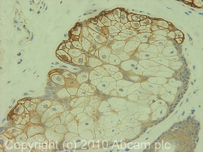
Anti-CD166 antibody (ab78649)
Key features and details
- Rabbit polyclonal to CD166
- Suitable for: IHC-P
- Reacts with: Human
- Isotype: IgG
Overview
-
Product name
Anti-CD166 antibody
See all CD166 primary antibodies -
Description
Rabbit polyclonal to CD166 -
Host species
Rabbit -
Tested Applications & Species
See all applications and species dataApplication Species IHC-P Human -
Immunogen
Synthetic peptide corresponding to Human CD166 aa 100-200 conjugated to keyhole limpet haemocyanin.
(Peptide available asab104421) -
Positive control
- This antibody gave a positive signal in Human normal skin FFPE section.
Properties
-
Form
Liquid -
Storage instructions
Shipped at 4°C. Store at +4°C short term (1-2 weeks). Upon delivery aliquot. Store at -20°C or -80°C. Avoid freeze / thaw cycle. -
Storage buffer
pH: 7.40
Preservative: 0.02% Sodium azide
Constituent: PBS
Batches of this product that have a concentration Concentration information loading...
Purity
Immunogen affinity purifiedClonality
PolyclonalIsotype
IgGResearch areas
Associated products
-
Compatible Secondaries
-
Isotype control
-
Recombinant Protein
Applications
The Abpromise guarantee
Our Abpromise guarantee covers the use of ab78649 in the following tested applications.
The application notes include recommended starting dilutions; optimal dilutions/concentrations should be determined by the end user.
GuaranteedTested applications are guaranteed to work and covered by our Abpromise guarantee.
PredictedPredicted to work for this combination of applications and species but not guaranteed.
IncompatibleDoes not work for this combination of applications and species.
Application Species IHC-P HumanAll applications RabbitCowDogPigOrangutanApplication Abreviews Notes IHC-P (1) Use a concentration of 1 µg/ml. Perform heat mediated antigen retrieval with citrate buffer pH 6 before commencing with IHC staining protocol.Notes IHC-P
Use a concentration of 1 µg/ml. Perform heat mediated antigen retrieval with citrate buffer pH 6 before commencing with IHC staining protocol.Target
-
Function
Cell adhesion molecule that binds to CD6. Involved in neurite extension by neurons via heterophilic and homophilic interactions. May play a role in the binding of T- and B-cells to activated leukocytes, as well as in interactions between cells of the nervous system. -
Tissue specificity
Spleen, placenta, liver, and weakly in liver. Expressed by activated T-cells, B-cells, monocytes and thymic epithelial cells. Expressed by neurons in the brain. Restricted expression in tumor cell lines. Preferentially expressed in highly metastasizing melanoma cell lines. -
Sequence similarities
Contains 3 Ig-like C2-type (immunoglobulin-like) domains.
Contains 2 Ig-like V-type (immunoglobulin-like) domains. -
Domain
The CD6 binding site is located in the N-terminal Ig-like domain. -
Cellular localization
Membrane. - Information by UniProt
-
Database links
- Entrez Gene: 281614 Cow
- Entrez Gene: 478550 Dog
- Entrez Gene: 214 Human
- Entrez Gene: 397269 Pig
- Entrez Gene: 100009305 Rabbit
- Omim: 601662 Human
- SwissProt: Q9BH13 Cow
- SwissProt: O46634 Dog
see all -
Alternative names
- Activated leukocyte cell adhesion molecule antibody
- ALCAM antibody
- ALCAM protein antibody
see all
Images
-
 Immunohistochemistry (Formalin/PFA-fixed paraffin-embedded sections) - Anti-CD166 antibody (ab78649)IHC image of ab78649 staining in human normal skin FFPE section, performed on a BondTM system using the standard protocol F. The section was pre-treated using heat mediated antigen retrieval with sodium citrate buffer (pH6, epitope retrieval solution 1) for 20 mins. The section was then incubated with ab78649, 1µg/ml for 15 mins at room temperature and detected using an HRP conjugated compact polymer system. DAB was used as the chromogen. The section was then counterstained with haematoxylin and mounted with DPX
Datasheets and documents
References (2)
ab78649 has been referenced in 2 publications.
- Rodríguez-Barrientos CA et al. Arresting proliferation improves the cell identity of corneal endothelial cells in the New Zealand rabbit. Mol Vis 25:745-755 (2019). PubMed: 31814700
- Landa-Solís C et al. Cryopreserved CD90+ cells obtained from mobilized peripheral blood in sheep: a new source of mesenchymal stem cells for preclinical applications. Cell Tissue Bank : (2015). IF . PubMed: 26220398
Images
-
 Immunohistochemistry (Formalin/PFA-fixed paraffin-embedded sections) - Anti-CD166 antibody (ab78649)IHC image of ab78649 staining in human normal skin FFPE section, performed on a BondTM system using the standard protocol F. The section was pre-treated using heat mediated antigen retrieval with sodium citrate buffer (pH6, epitope retrieval solution 1) for 20 mins. The section was then incubated with ab78649, 1µg/ml for 15 mins at room temperature and detected using an HRP conjugated compact polymer system. DAB was used as the chromogen. The section was then counterstained with haematoxylin and mounted with DPX


