Anti-ACE2 antibody (ab15348)
Key features and details
- Rabbit polyclonal to ACE2
- Suitable for: IHC-Fr, IHC (PFA fixed), WB, IHC-P
- Reacts with: Mouse, Rat, Human
- Isotype: IgG
Overview
-
Product name
Anti-ACE2 antibody
See all ACE2 primary antibodies -
Description
Rabbit polyclonal to ACE2 -
Host species
Rabbit -
Specificity
No cross reactivity to ACE1.
-
Tested Applications & Species
See all applications and species dataApplication Species IHC (PFA fixed) MouseRatIHC-Fr HumanIHC-P HumanWB MouseRatHuman -
Immunogen
Synthetic peptide within Human ACE2 aa 750 to the C-terminus. The exact immunogen sequence used to generate this antibody is proprietary information. If additional detail on the immunogen is needed to determine the suitability of the antibody for your needs, please contact our Scientific Support team to discuss your requirements.
Database link: Q9BYF1
(Peptide available asab15352)
Properties
-
Form
Liquid -
Storage instructions
Shipped at 4°C. Store at +4°C short term (1-2 weeks). Upon delivery aliquot. Store at -20°C long term. Avoid freeze / thaw cycle. -
Storage buffer
pH: 7.2
Preservative: 0.02% Sodium azide -
 Concentration information loading...
 -
Purity
Immunogen affinity purified -
Primary antibody notes
No cross reactivity to ACE1. -
Clonality
Polyclonal -
Isotype
IgG -
Research areas
Images
-
All lanes : Anti-ACE2 antibody (ab15348) at 2 µg/ml
Lane 1 : Human testis tissue lysate
Lane 2 : Human lung tissue lysate
Lane 3 : Human intestine tissue lysate
Lane 4 : Human breast tissue lysate
Lane 5 : Caco2 cell lysateLysates/proteins at 15 µg per lane.
Secondary
All lanes : Goat anti-rabbit IgG HRP conjugate at 1/10000 dilution
Predicted band size: 97 kDa -
Immunohistochemistry (Formalin/PFA-fixed paraffin-embedded sections) analysis of human lung tissue labeling ACE2 with ab15348 at 20 μg/mL.
Goat anti-rabbit IgG secondary antibody at 1/500 dilution (green) and DAPI staining (blue).
-
Immunohistochemistry (Formalin/PFA-fixed paraffin-embedded sections) analysis of human testis tissue labeling ACE2 with ab15348 at 20 μg/mL.
Goat anti-rabbit IgG secondary antibody at 1/500 dilution (green) and DAPI staining (blue).
-
All lanes : Anti-ACE2 antibody (ab15348) at 2 µg/ml
Lane 1 : Rat skin tissue lysate
Lane 2 : Rat spleen tissue lysate
Lane 3 : Rat heart tissue lysate
Lane 4 : Rat brain tissue lysate
Lysates/proteins at 15 µg per lane.
Secondary
All lanes : Goat anti-rabbit IgG HRP conjugate at 1/10000 dilution
Predicted band size: 97 kDaWestern blot of ab15348 incubated for 1h at RT in 5% NFDM/TBST.
-
All lanes : Anti-ACE2 antibody (ab15348) at 2 µg/ml
Lane 1 : Mouse liver tissue lysate
Lane 2 : Mouse spleen tissue lysate
Lane 3 : Mouse heart tissue lysate
Lane 4 : Mouse bladder tissue lysate
Lane 5 : Mouse pancreas tissue lysate
Lane 6 : Mouse stomach tissue lysate
Lysates/proteins at 15 µg per lane.
Secondary
All lanes : Goat anti-rabbit IgG HRP conjugate at 1/10000 dilution
Predicted band size: 97 kDaWestern blot of ab15348 incubated for 1h at RT in 5% NFDM/TBST.
-
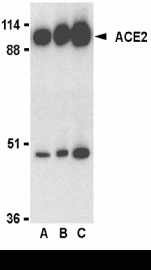
Western blot analysis of ACE2 in human kidney lysate with ab15438 at 0.5 μg/mL (lane A), 1 μg/mL (lane B), and 2 μg/mL (lane C).
-
Immunohistochemistry (Formalin/PFA-fixed paraffin-embedded sections) analysis of mouse lung tissue labeling ACE2 with ab15348 at 20 μg/mL.
Goat anti-rabbit IgG secondary antibody at 1/500 dilution (green) and DAPI staining (blue).
-
Immunohistochemistry (Formalin/PFA-fixed paraffin-embedded sections) analysis of rat lung tissue labeling ACE2 with ab15348 at 20 μg/mL.
Goat anti-rabbit IgG secondary antibody at 1/500 dilution (green) and DAPI staining (blue).
-
Immunohistochemistry (Formalin/PFA-fixed paraffin-embedded sections) analysis of human kidney tissue labeling ACE2 with ab15348 at 2 μg/mL.
Tissue was fixed with formaldehyde and blocked with 10% serum for 1 hour at room temperature; antigen retrieval was by heat mediation with a citrate buffer (pH6). Samples were incubated with primary antibody overnight at 4°C. A goat anti-rabbit IgG HRP at 1/250 was used as secondary. Counter stained with Hematoxylin.


