Anti-Caveolin-3 antibody - Caveolae Marker (ab2912)
")
Key features and details
- Rabbit polyclonal to Caveolin-3 - Caveolae Marker
- Suitable for: IHC-P, ICC/IF, Flow Cyt, WB, IP
- Reacts with: Mouse, Rat, Human
- Isotype: IgG
Overview
-
Product name
Anti-Caveolin-3 antibody - Caveolae Marker
See all Caveolin-3 primary antibodies -
Description
Rabbit polyclonal to Caveolin-3 - Caveolae Marker -
Host species
Rabbit -
Specificity
This antibody does not detect caveolin-1 or -2. -
Tested Applications & Species
See all applications and species dataApplication Species Flow Cyt HumanICC/IF HumanIHC-P MouseIP MouseWB MouseRatHuman
-
Immunogen
Synthetic peptide corresponding to Mouse Caveolin-3 aa 1-19.
Sequence:MMTEEHTDLEARIIKDIHC
(Peptide available asab4930)


Properties
-
Form
Liquid -
Storage instructions
Shipped at 4°C. Store at +4°C short term (1-2 weeks). Upon delivery aliquot. Store at -20°C long term. Avoid freeze / thaw cycle. -
Storage buffer
Preservative: 0.05% Sodium azide
Constituents: 0.1% BSA, 99% PBS -
 Concentration information loading...
 -
Purity
Immunogen affinity purified -
Purification notes
Antigen affinity chromatography. -
Clonality
Polyclonal -
Isotype
IgG -
Research areas
Images
-
Western blot - Anti-Caveolin-3 antibody - Caveolae Marker (ab2912)All lanes : Anti-Caveolin-3 antibody - Caveolae Marker (ab2912)
Lane 1 : Rat heart tissue lysate
Lane 2 : Mouse heart tissue lysate
Lane 3 : HEK293 cell lysate
Lane 4 : Rat skeletal muscle tissue lysate
Lane 5 : Mouse muscle tissue lysate
Lysates/proteins at 20 µg per lane.
Secondary
All lanes : HRP-conjugated goat anti-rabbit IgG (H+L) at 1/2500 dilution
Developed using the ECL technique.
Observed band size: 17 kDa why is the actual band size different from the predicted?Blocked with 5% skimmed milk.
-
 Immunocytochemistry/ Immunofluorescence - Anti-Caveolin-3 antibody - Caveolae Marker (ab2912)
Immunocytochemistry/Immunofluorescence analysis of Caveolin-3 in HeLa Cells. Cells were grown on chamber slides and fixed with formaldehyde prior to staining. Cells were probed without (control) (right panel) or with ab2912 at a dilution of 1/20 overnight at 4°C, washed with PBS and incubated with a DyLight-488 conjugated secondary antibody. Caveolin-3 staining (green), F-Actin staining with Phalloidin (red) and nuclei with DAPI (blue) is shown. Images were taken at 60X magnification.
-
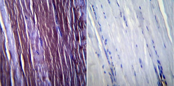
 Immunohistochemistry (Formalin/PFA-fixed paraffin-embedded sections) - Anti-Caveolin-3 antibody - Caveolae Marker (ab2912)
Immunohistochemistry was performed on normal biopsies of deparaffinized mouse heart tissue. To expose target proteins heat induced antigen retrieval was performed using 10mM sodium citrate (pH6.0) buffer microwaved for 8-15 minutes. Following antigen retrieval tissues were blocked in 3% BSA-PBS for 30 minutes at room temperature. Tissues were then probed at a dilution of 1/200 with a rabbit polyclonal antibody recognizing Caveolin-3 ab2912 or without primary antibody (negative control) overnight at 4°C in a humidified chamber. Tissues were washed extensively with PBST and endogenous peroxidase activity was quenched with a peroxidase suppressor. Detection was performed using a biotin-conjugated secondary antibody and SA-HRP followed by colorimetric detection using DAB. Tissues were counterstained with hematoxylin and prepped for mounting.
-
 Immunoprecipitation - Anti-Caveolin-3 antibody - Caveolae Marker (ab2912)
Caveolin-3 was immunoprecipitated using 5 µg of ab2912 from mouse heart tissue lysate (Lane 3) using the protein A beads. Normal rabbit IgG was used as a isotype control (Lane 2). 10% input represents the cell extract used for immunoprecipitation (Lane 1). Western blot analysis was performed using ab2912 and HRP-conjugated goat anti-rabbit IgG (H+L) at a dilution of 1/2500. Chemiluminescent detection was performed.
-
 Flow Cytometry - Anti-Caveolin-3 antibody - Caveolae Marker (ab2912)
Flow cytometry analysis of U-87 MG cells. Cells were fixed with 70% ethanol for 10 minutes, permeabilized with 0.25% Triton X-100 for 20 minutes, and blocked with 5% BSA for 30 minutes at room temperature. Cells were labeled with ab2912 (red histogram) or with rabbit isotype control (pink histogram) at 3-5 ug/million cells in 2.5% BSA. After incubation at room temperature for 2 hours, the cells were labeled with Alexa Fluor® 488-conjugated goat anti-rabbit secondary antibody at a dilution of 1/400 for 30 minutes at room temperature. The purple histogram represents unstained control cells and the green histogram represents no-primary-antibody control.
-
 Immunocytochemistry/ Immunofluorescence - Anti-Caveolin-3 antibody - Caveolae Marker (ab2912)
Immunofluorescent analysis of Caveolin-3 in C2C11 Cells. Cells were grown on chamber slides and fixed with formaldehyde prior to staining. Cells were probed without (control) (right panel) or with ab2912 at a dilution of 1/20 overnight at 4 C, washed with PBS and incubated with a DyLight-488 conjugated secondary antibody. Caveolin-3 staining (green), F-Actin staining with Phalloidin (red) and nuclei with DAPI (blue) is shown. Images were taken at 60X magnification.
-
 Immunohistochemistry (Formalin/PFA-fixed paraffin-embedded sections) - Anti-Caveolin-3 antibody - Caveolae Marker (ab2912)
Immunohistochemistry was performed on normal biopsies of deparaffinized Mouse skeletal muscle tissue. To expose target proteins heat induced antigen retrieval was performed using 10mM sodium citrate (pH6.0) buffer microwaved for 8-15 minutes. Following antigen retrieval tissues were blocked in 3% BSA-PBS for 30 minutes at room temperature. Tissues were then probed at a dilution of 1/100 with a rabbit polyclonal antibody recognizing Caveolin-3 ab2912 or without primary antibody (negative control) overnight at 4°C in a humidified chamber. Tissues were washed extensively with PBST and endogenous peroxidase activity was quenched with a peroxidase suppressor. Detection was performed using a biotin-conjugated secondary antibody and SA-HRP followed by colorimetric detection using DAB. Tissues were counterstained with hematoxylin and prepped for mounting.
-
 Western blot - Anti-Caveolin-3 antibody - Caveolae Marker (ab2912)Anti-Caveolin-3 antibody - Caveolae Marker (ab2912) + Rat cardiac muscle tissue lysate
-
 Immunohistochemistry (Formalin/PFA-fixed paraffin-embedded sections) - Anti-Caveolin-3 antibody - Caveolae Marker (ab2912)
Immunohistochemistry was performed on normal biopsies of deparaffinized mouse lymph node tissue. To expose target proteins heat induced antigen retrieval was performed using 10mM sodium citrate (pH6.0) buffer microwaved for 8-15 minutes. Following antigen retrieval tissues were blocked in 3% BSA-PBS for 30 minutes at room temperature. Tissues were then probed at a dilution of 1/200 with a rabbit polyclonal antibody recognizing Caveolin-3 ab2912 or without primary antibody (negative control) overnight at 4°C in a humidified chamber. Tissues were washed extensively with PBST and endogenous peroxidase activity was quenched with a peroxidase suppressor. Detection was performed using a biotin-conjugated secondary antibody and SA-HRP followed by colorimetric detection using DAB. Tissues were counterstained with hematoxylin and prepped for mounting.
-
 Immunocytochemistry/ Immunofluorescence - Anti-Caveolin-3 antibody - Caveolae Marker (ab2912)
Immunocytochemistry/Immunofluorescence analysis of 70% confluent log phase A-375 cells. Cells were fixed with 4% paraformaldehyde for 15 minutes, permeabilized with 0.25% Triton X-100 for 10 minutes, and blocked with 5% BSA for 1 hour at room temperature. Samples were incubated with ab2912 at 1µg/ml in 1% BSA for 3 hours at room temperature and then labelled with Alexa Fluor® 488-conjugated goat anti-rabbit IgG (H+L) at a dilution of 1/2000 for 45 minutes at room temperature (panel a: green). Nuclei (panel b: blue) were stained with DAPI. F-actin (panel c: red) was stained with Alexa Fluor® 555 Rhodamine Phalloidin (1/300). Panel d is a merged image showing cytoplasmic localization. Panel e is a no primary antibody control. The images were captured at 60X magnification.


