Anti-Catalase antibody - Peroxisome Marker (ab16731)
")
Key features and details
- Rabbit polyclonal to Catalase - Peroxisome Marker
- Suitable for: ICC/IF, IHC-P, WB
- Reacts with: Mouse, Rat, Human
- Isotype: IgG
Overview
-
Product name
Anti-Catalase antibody - Peroxisome Marker
See all Catalase primary antibodies -
Description
Rabbit polyclonal to Catalase - Peroxisome Marker -
Host species
Rabbit -
Tested Applications & Species
See all applications and species dataApplication Species ICC/IF HumanIHC-P HumanWB Human
-
Immunogen
Recombinant human protein purified from E.coli
Properties
-
Form
Liquid -
Storage instructions
Shipped at 4°C. Store at +4°C short term (1-2 weeks). Upon delivery aliquot. Store at -20°C. Avoid freeze / thaw cycle. -
Storage buffer
Preservative: 0.03% Sodium azide
Constituents: HEPES, 50% Glycerol, 0.87% Sodium chloride, 0.01% BSA -
 Concentration information loading...
 -
Purity
Protein A purified -
Clonality
Polyclonal -
Isotype
IgG -
Research areas
Images
-
Western blot - Anti-Catalase antibody - Peroxisome Marker (ab16731)
Western Blot analysis of cell lysates.
Lane 1: HeLa cell lysates
Lane 2: Jurkat cell lysates
Lane 3: Mouse brain
Lane 4: Rat brainThe band marked with NS is probably non-specific.
-
 Immunocytochemistry/ Immunofluorescence - Anti-Catalase antibody - Peroxisome Marker (ab16731)ICC/IF image of ab16731 stained Hela cells. The cells were 4% PFA fixed (10 min) and then incubated in 1%BSA / 10% normal goat serum / 0.3M glycine in 0.1% PBS-Tween for 1h to permeabilise the cells and block non-specific protein-protein interactions. The cells were then incubated with the antibody (ab16731, 5µg/ml) overnight at +4°C. The secondary antibody (green) was DyLight® 488 goat anti-rabbit IgG - H&L, pre-adsorbed (ab96899) used at a 1/250 dilution for 1h. Alexa Fluor® 594 WGA was used to label plasma membranes (red) at a 1/200 dilution for 1h. DAPI was used to stain the cell nuclei (blue) at a concentration of 1.43µM.
-
 Immunohistochemistry (Formalin/PFA-fixed paraffin-embedded sections) - Anti-Catalase antibody - Peroxisome Marker (ab16731)Ab16731 staining human normal adrenal gland tissue. Staining is localised to intracellular compartment (peroxisomes).
Left panel: with primary antibody at 1 ug/ml. Right panel: isotype control.
Sections were stained using an automated system DAKO Autostainer Plus , at room temperature. Sections were rehydrated and antigen retrieved with the Dako 3-in-1 AR buffer EDTA pH 9.0 in a DAKO PT Link. Slides were peroxidase blocked in 3% H2O2 in methanol for 10 minutes. They were then blocked with Dako Protein block for 10 minutes (containing casein 0.25% in PBS) then incubated with primary antibody for 20 minutes and detected with Dako Envision Flex amplification kit for 30 minutes. Colorimetric detection was completed with diaminobenzidine for 5 minutes. Slides were counterstained with Haematoxylin and coverslipped under DePeX. Please note that for manual staining we recommend to optimize the primary antibody concentration and incubation time (overnight incubation), and amplification -
 Western blot - Anti-Catalase antibody - Peroxisome Marker (ab16731)All lanes : Anti-Catalase antibody - Peroxisome Marker (ab16731) at 1/2000 dilution
Lane 1 : 40ug supernatant of mouse liver homogenate
Lane 2 : 20ug supernatant of mouse liver homogenate
Lane 3 : 5ug supernatant of mouse liver homogenate
Secondary
All lanes : HRP conjugated donkey anti-rabbit antibody
Developed using the ECL technique.
Performed under reducing conditions.
Predicted band size: 60 kDa
Observed band size: 60 kDa
Exposure time: 1 minute
This image is courtesy of an Abreview submitted by Sandra Sobocanec on 16 March 2006. -
 Immunocytochemistry/ Immunofluorescence - Anti-Catalase antibody - Peroxisome Marker (ab16731) This image is a courtesy of an anonymous Abreview.
ab16731 at 1/200 dilution staining Catalase in human 293FT cells by Immunocytochemistry/ Immunofluorescence. Cells were fixed in formaldehyde and blocked in 5% BSA for 1 hour at 25°C. The primary antibody was used at 1/200 dilution in PBS and incubated with sample at 4°C for 12 hours. An Alexa Fluor® 488 conjugated Goat polyclonal to rabbit IgG was used undiluted as secondary.
-
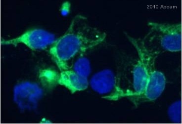
 Immunocytochemistry/ Immunofluorescence - Anti-Catalase antibody - Peroxisome Marker (ab16731) This image is courtesy of an anonymous abreview.ab16731 at a 1/200 dilution staining Catalase in mouse bone marrow cells by Immunocytochemistry/ Immunofluorescence, incubated for 9 hours at 4°C. Formalin fixed. Blocked with 2% BSA for 30 minutes at 20°C. Secondary used at 1/200 dilution polyclonal Goat anti-rabbit IgG conjugated to Alexa Fluor 488 (green). Nuclei stained with DAPI (blue).


