Anti-Casein Kinase 1 delta/CSNK1D antibody (ab236601)
")
Key features and details
- Rabbit polyclonal to Casein Kinase 1 delta/CSNK1D
- Suitable for: IP, ICC/IF, IHC-P, WB
- Reacts with: Human
- Isotype: IgG
Overview
-
Product name
Anti-Casein Kinase 1 delta/CSNK1D antibody
See all Casein Kinase 1 delta/CSNK1D primary antibodies -
Description
Rabbit polyclonal to Casein Kinase 1 delta/CSNK1D -
Host species
Rabbit -
Tested applications
Suitable for: IP, ICC/IF, IHC-P, WBmore details -
Species reactivity
Reacts with: Human
Predicted to work with: Mouse, Rat, Cow, Xenopus laevis, Zebrafish, Orangutan, Xenopus tropicalis
-
Immunogen
Recombinant fragment corresponding to Human Casein Kinase 1 delta/CSNK1D aa 150-400.
Database link: P48730 -
Positive control
- WB: HeLa whole cell lysate. IHC-P: Human skeletal muscle, liver cancer and lymph node tissue. ICC/IF: HepG2 cells. IP: HeLa whole cell lysate.
-
General notes
Previously labelled as Casein Kinase 1 delta.
The Life Science industry has been in the grips of a reproducibility crisis for a number of years. Abcam is leading the way in addressing the problem with our range of recombinant monoclonal antibodies and knockout edited cell lines for gold-standard validation.
One factor contributing to the crisis is the use of antibodies that are not suitable. This can lead to misleading results and the use of incorrect data informing project assumptions and direction. To help address this challenge, we have introduced an application and species grid on our primary antibody datasheets to make it easy to simplify identification of the right antibody for your needs.
Learn more here.


Properties
-
Form
Liquid -
Storage instructions
Shipped at 4°C. Store at +4°C short term (1-2 weeks). Upon delivery aliquot. Store at -20°C long term. Avoid freeze / thaw cycle. -
Storage buffer
pH: 7.40
Constituents: 50% Glycerol (glycerin, glycerine), PBS, 0.03% Proclin 300 -
 Concentration information loading...
 -
Purity
Protein G purified -
Purification notes
Purity >95%. -
Clonality
Polyclonal -
Isotype
IgG -
Research areas
Images
-
Western blot - Anti-Casein Kinase 1 delta/CSNK1D antibody (ab236601)Anti-Casein Kinase 1 delta/CSNK1D antibody (ab236601) at 1/1000 dilution + HeLa (Human epithelial cell line from cervix adenocarcinoma) whole cell lysate
Secondary
Goat polyclonal to rabbit IgG at 1/50000 dilution
-
 Immunohistochemistry (Formalin/PFA-fixed paraffin-embedded sections) - Anti-Casein Kinase 1 delta/CSNK1D antibody (ab236601)
Paraffin-embedded human skeletal muscle tissue stained for Casein Kinase 1 delta/CSNK1D with ab236601 at a 1/1000 dilution in immunohistochemical analysis.
After dewaxing and hydration, antigen retrieval was mediated by high pressure in a citrate buffer (pH 6.0). Section was blocked with 10% normal goat serum 30 minutes at RT. Then primary antibody (1% BSA) was incubated at 4°C overnight. The primary is detected by a biotinylated secondary antibody and visualized tissue using an HRP conjugated SP system.
-
 Immunocytochemistry/ Immunofluorescence - Anti-Casein Kinase 1 delta/CSNK1D antibody (ab236601)
HepG2 (Human liver hepatocellular carcinoma cell line) cells stained for Casein Kinase 1 delta/CSNK1D using ab236601 at a dilution of 1/333 in ICC/IF.
The cells were fixed in 4% formaldehyde, permeabilized using 0.2% Triton X-100 and blocked in 10% normal goat serum. The cells were then incubated with the primary antibody overnight at 4°C. Secondary used is an Alexa-Fluor®488-conjugated Goat Anti-Rabbit IgG (H+L).
-
 Immunoprecipitation - Anti-Casein Kinase 1 delta/CSNK1D antibody (ab236601)
Casein Kinase 1 delta/CSNK1D was immunoprecipitated from HeLa (Human epithelial cell line from cervix adenocarcinoma) whole cell lysate using 6 µg of ab236601.
Lane 1: Rabbit control IgG (1 µg) in HeLa whole cell lysate.
Lane 2: ab236601 (6 µg) in HeLa whole cell lysate.
Lane 3: HeLa whole cell lysate (10 µg input).
For western blotting an HRP-conjugated Protein G antibody was used as the secondary antibody at a 1/2000 dilution.
-
 Immunohistochemistry (Formalin/PFA-fixed paraffin-embedded sections) - Anti-Casein Kinase 1 delta/CSNK1D antibody (ab236601)
Paraffin-embedded human liver cancer tissue stained for Casein Kinase 1 delta/CSNK1D with ab236601 at a 1/1000 dilution in immunohistochemical analysis.
After dewaxing and hydration, antigen retrieval was mediated by high pressure in a citrate buffer (pH 6.0). Section was blocked with 10% normal goat serum 30 minutes at RT. Then primary antibody (1% BSA) was incubated at 4°C overnight. The primary is detected by a biotinylated secondary antibody and visualized tissue using an HRP conjugated SP system.
-
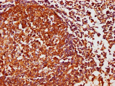 Immunohistochemistry (Formalin/PFA-fixed paraffin-embedded sections) - Anti-Casein Kinase 1 delta/CSNK1D antibody (ab236601)
Paraffin-embedded human lymph node tissue stained for Casein Kinase 1 delta/CSNK1D with ab236601 at a 1/1000 dilution in immunohistochemical analysis.
After dewaxing and hydration, antigen retrieval was mediated by high pressure in a citrate buffer (pH 6.0). Section was blocked with 10% normal goat serum 30 minutes at RT. Then primary antibody (1% BSA) was incubated at 4°C overnight. The primary is detected by a biotinylated secondary antibody and visualized tissue using an HRP conjugated SP system.


