Anti-Aspartyl Aminopeptidase antibody (ab239031)
")
Key features and details
- Rabbit polyclonal to Aspartyl Aminopeptidase
- Suitable for: IHC-P, ICC/IF
- Reacts with: Human
- Isotype: IgG
Overview
-
Product name
Anti-Aspartyl Aminopeptidase antibody
See all Aspartyl Aminopeptidase primary antibodies -
Description
Rabbit polyclonal to Aspartyl Aminopeptidase -
Host species
Rabbit -
Tested Applications & Species
See all applications and species dataApplication Species ICC/IF HumanIHC-P Human
-
Immunogen
Recombinant fragment corresponding to Human Aspartyl Aminopeptidase aa 250-400.
Database link: Q9ULA0 -
Positive control
- IHC-P: Human small intestine tissue. ICC/IF: MCF7 cells.


Properties
-
Form
Liquid -
Storage instructions
Shipped at 4°C. Store at +4°C short term (1-2 weeks). Upon delivery aliquot. Store at -20°C long term. Avoid freeze / thaw cycle. -
Storage buffer
pH: 7.40
Constituents: 50% Glycerol (glycerin, glycerine), PBS, 0.03% Proclin 300 -
 Concentration information loading...
 -
Purity
Protein G purified -
Purification notes
Purity greater than 95%. -
Clonality
Polyclonal -
Isotype
IgG -
Research areas
Images
-
Immunocytochemistry/ Immunofluorescence - Anti-Aspartyl Aminopeptidase antibody (ab239031)
MCF7 (Human breast adenocarcinoma cell line) cells stained for Aspartyl Aminopeptidase (Green) using ab239031 at 1/200 dilution in ICC/IF, followed by an Alexa-Fluor®488-conjugated Goat Anti-Rabbit IgG (H+L) secondary antibody.
The cells were fixed in 4% formaldehyde, permeabilized using 0.2% Triton X-100 and blocked in 10% normal goat serum. The cells were then incubated with the antibody overnight at 4°C. The nuclear counterstain was DAPI (Blue).
-
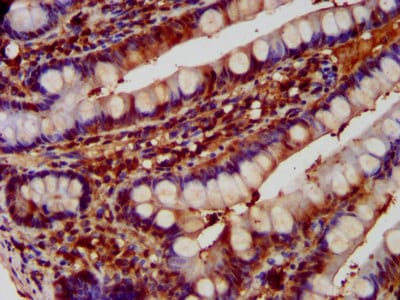 Immunohistochemistry (Formalin/PFA-fixed paraffin-embedded sections) - Anti-Aspartyl Aminopeptidase antibody (ab239031)
Paraffin-embedded human small intestine tissue stained for Aspartyl Aminopeptidase, using ab239031 at 1/600 dilution in immunohistochemical analysis.
After dewaxing and hydration, antigen retrieval was mediated by high pressure in a citrate buffer (pH 6.0). Section was blocked with 10% normal goat serum 30 minutes at RT. Then primary antibody (1% BSA) was incubated at 4°C overnight. The primary is detected by a biotinylated secondary antibody and visualized using an HRP conjugated SP system.


