Anti-ALR/HPO antibody (ab36376)
")
Key features and details
- Rabbit polyclonal to ALR/HPO
- Suitable for: WB, ICC/IF
- Reacts with: Mouse, Human
- Isotype: IgG
Overview
-
Product name
Anti-ALR/HPO antibody
See all ALR/HPO primary antibodies -
Description
Rabbit polyclonal to ALR/HPO -
Host species
Rabbit -
Tested applications
Suitable for: WB, ICC/IFmore details -
Species reactivity
Reacts with: Mouse, Human
Predicted to work with: Rat, Xenopus laevis
-
Immunogen
-
Positive control
- Purchase matching WB positive control:Recombinant Human ALR/HPO protein
- Recombinant Human ALR/HPO protein (ab114811) can be used as a positive control in WB. This antibody gave a positive signal in mouse liver and mouse testis tissue lysates.
-
General notes
This product was previously labelled as ALR
Properties
-
Form
Liquid -
Storage instructions
Shipped at 4°C. Store at +4°C short term (1-2 weeks). Upon delivery aliquot. Store at -20°C or -80°C. Avoid freeze / thaw cycle. -
Storage buffer
pH: 7.40
Preservative: 0.02% Sodium azide
Constituent: PBS
Batches of this product that have a concentration Concentration information loading...
Purity
Immunogen affinity purifiedClonality
PolyclonalIsotype
IgGResearch areas
Associated products
-
Compatible Secondaries
-
Isotype control
Applications
Our Abpromise guarantee covers the use of ab36376 in the following tested applications.
The application notes include recommended starting dilutions; optimal dilutions/concentrations should be determined by the end user.
Application Abreviews Notes WB Use a concentration of 1 µg/ml. Detects a band of approximately 23 kDa (predicted molecular weight: 23 kDa). ICC/IF Use a concentration of 1 µg/ml. Target
-
Function
Isoform 1: FAD-dependent sulfhydryl oxidase. Within the mitochondrial intermembrane space, participates in a chain of disulfide exchange reactions with MIA40, that generate disulfide bonds in a number of resident proteins with twin Cx3C and Cx9C motifs.
Isoform 2: May act as an autocrine hepatotrophic growth factor promoting liver regeneration. -
Tissue specificity
Ubiquitously expressed. Highest expression in the testis and liver and low expression in the muscle. -
Involvement in disease
Defects in GFER are a cause of mitochondrial progressive myopathy with congenital cataract hearing loss and developmental delay (MPMCHD) [MIM:613076]; also called combined mitochondrial complex deficiency. -
Sequence similarities
Contains 1 ERV/ALR sulfhydryl oxidase domain. -
Cellular localization
Mitochondrion intermembrane space and Cytoplasm. Secreted. - Information by UniProt
-
Database links
- Entrez Gene: 2671 Human
- Entrez Gene: 11692 Mouse
- Entrez Gene: 27100 Rat
- Omim: 600924 Human
- SwissProt: P55789 Human
- SwissProt: P56213 Mouse
- SwissProt: Q63042 Rat
- Unigene: 726325 Human
see all -
Alternative names
- ALR_HUMAN antibody
- Augmenter of liver regeneration antibody
- ERV1 antibody
see all
Images
-
Western blot - Anti-ALR/HPO antibody (ab36376)All lanes : Anti-ALR/HPO antibody (ab36376) at 1 µg/ml
Lane 1 : Liver (Mouse) Tissue Lysate
Lane 2 : Testis (Mouse) Tissue Lysate
Lysates/proteins at 10 µg per lane.
Secondary
All lanes : Goat Anti-Rabbit IgG H&L (HRP) preadsorbed (ab97080) at 1/5000 dilution
Predicted band size: 23 kDa
Observed band size: 23 kDa
Abcam recommends using milk as the blocking agent. Abcam welcomes customer feedback and would appreciate any comments regarding this product and the data presented above. -
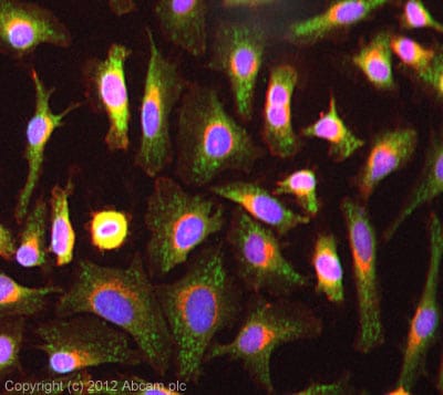 Immunocytochemistry/ Immunofluorescence - Anti-ALR/HPO antibody (ab36376)ICC/IF image of ab36376 stained HeLa cells. The cells were 100% methanol fixed (5 min) and then incubated in 1%BSA / 10% normal goat serum / 0.3M glycine in 0.1% PBS-Tween for 1h to permeabilise the cells and block non-specific protein-protein interactions. The cells were then incubated with the antibody (ab36376, 5µg/ml) overnight at +4°C. The secondary antibody (green) was ab96899, DyLight® 488 goat anti-rabbit IgG (H+L) used at a 1/250 dilution for 1h. Alexa Fluor® 594 WGA was used to label plasma membranes (red) at a 1/200 dilution for 1h. DAPI was used to stain the cell nuclei (blue) at a concentration of 1.43µM. This antibody also gave a positive result in 4% PFA fixed (10 min) HepG2 and MCF7 cells at 5µg/ml, and in 100% methanol fixed (5 min) MCF7 cells at 5µg/ml.
Protocols
Datasheets and documents
References (0)
ab36376 has not yet been referenced specifically in any publications.
Images
-
Western blot - Anti-ALR/HPO antibody (ab36376)All lanes : Anti-ALR/HPO antibody (ab36376) at 1 µg/ml
Lane 1 : Liver (Mouse) Tissue Lysate
Lane 2 : Testis (Mouse) Tissue Lysate
Lysates/proteins at 10 µg per lane.
Secondary
All lanes : Goat Anti-Rabbit IgG H&L (HRP) preadsorbed (ab97080) at 1/5000 dilution
Predicted band size: 23 kDa
Observed band size: 23 kDa
Abcam recommends using milk as the blocking agent. Abcam welcomes customer feedback and would appreciate any comments regarding this product and the data presented above. -
Immunocytochemistry/ Immunofluorescence - Anti-ALR/HPO antibody (ab36376)ICC/IF image of ab36376 stained HeLa cells. The cells were 100% methanol fixed (5 min) and then incubated in 1%BSA / 10% normal goat serum / 0.3M glycine in 0.1% PBS-Tween for 1h to permeabilise the cells and block non-specific protein-protein interactions. The cells were then incubated with the antibody (ab36376, 5µg/ml) overnight at +4°C. The secondary antibody (green) was ab96899, DyLight® 488 goat anti-rabbit IgG (H+L) used at a 1/250 dilution for 1h. Alexa Fluor® 594 WGA was used to label plasma membranes (red) at a 1/200 dilution for 1h. DAPI was used to stain the cell nuclei (blue) at a concentration of 1.43µM. This antibody also gave a positive result in 4% PFA fixed (10 min) HepG2 and MCF7 cells at 5µg/ml, and in 100% methanol fixed (5 min) MCF7 cells at 5µg/ml.


