Anti-ALDH1L1 antibody (ab235197)
")
Key features and details
- Goat polyclonal to ALDH1L1
- Suitable for: WB, ICC/IF, IHC-P
- Reacts with: Mouse, Human
- Isotype: IgG
Overview
-
Product name
Anti-ALDH1L1 antibody
See all ALDH1L1 primary antibodies -
Description
Goat polyclonal to ALDH1L1 -
Host species
Goat -
Tested applications
Suitable for: WB, ICC/IF, IHC-Pmore details -
Species reactivity
Reacts with: Mouse, Human
Predicted to work with: Rat, Orangutan
-
Immunogen
Synthetic peptide. This information is proprietary to Abcam and/or its suppliers.
-
Positive control
- WB: Recombinant mouse ALDH1L1 protein; Mouse liver tissue lysate. ICC/IF: NIH/3T3 cells. IHC-P: Mouse kidney and liver tissue.
-
General notes
Reproducibility is key to advancing scientific discovery and accelerating scientists’ next breakthrough.
Abcam is leading the way with our range of recombinant antibodies, knockout-validated antibodies and knockout cell lines, all of which support improved reproducibility.
We are also planning to innovate the way in which we present recommended applications and species on our product datasheets, so that only applications & species that have been tested in our own labs, our suppliers or by selected trusted collaborators are covered by our Abpromise™ guarantee.
In preparation for this, we have started to update the applications & species that this product is Abpromise guaranteed for.
We are also updating the applications & species that this product has been “predicted to work with,” however this information is not covered by our Abpromise guarantee.
Applications & species from publications and Abreviews that have not been tested in our own labs or in those of our suppliers are not covered by the Abpromise guarantee.
Please check that this product meets your needs before purchasing. If you have any questions, special requirements or concerns, please send us an inquiry and/or contact our Support team ahead of purchase. Recommended alternatives for this product can be found below, as well as customer reviews and Q&As.
Properties
-
Form
Liquid -
Storage instructions
Shipped at 4°C. Store at +4°C short term (1-2 weeks). Upon delivery aliquot. Store at -20°C. Avoid freeze / thaw cycle. -
Storage buffer
Preservative: 0.01% Sodium azide
Constituent: PBS -
 Concentration information loading...
 -
Purity
Affinity purified -
Clonality
Polyclonal -
Isotype
IgG -
Research areas
Images
-
Western blot - Anti-ALDH1L1 antibody (ab235197)All lanes : Anti-ALDH1L1 antibody (ab235197) at 1/1000 dilution
Lane 1 : Opal pre-stained ladder
Lane 2 : Recombinant mouse ALDH1L1 protein
Lane 3 : HEK-293T (human epithelial cell line from embryonic kidney transformed with large T antigen) cell lysate
Lane 4 : NIH/3T3 (mouse embryo fibroblast cell line) cell lysate
Lane 5 : Mouse liver (normal tissue) lysate
Secondary
Lanes 1-4 : F(ab)'2 Dk-a-Gt IgG [H&L] at 1/40000 dilution
Lane 5 : Goat polyclonal to ALDH1L1 (ab235197) at 1/40000 dilution (F(ab)'2 Dk-a-Gt IgG [H&L])
Predicted band size: 99 kDa
Observed band size: 98.7 kDa why is the actual band size different from the predicted?
Exposure time: 30 seconds
-
 Immunocytochemistry/ Immunofluorescence - Anti-ALDH1L1 antibody (ab235197)
4% paraformaldehyde-fixed, 0.3% Triton X-100 permeabilized NIH/3T3 (mouse embryo fibroblast cell line) cells stained for ALDH1L1 (Green) using ab235197 at 15 μg/mL over night at 4°C in ICC/IF. The nuclear counterstain is DAPI (Blue).
Donkey Anti-Goat IgG DyLight™ 488 Conjugated Preadsorbed was used as the secondary antibody at 5 μg/mL for 1 hour at room temperature.
A) DAPI
B) ab235197 + secondary.
C) Merged image
D) Secondary only control.Expected localization: Cytoplasm.
-
 Immunocytochemistry/ Immunofluorescence - Anti-ALDH1L1 antibody (ab235197)
100% methanol-fixed, 0.3% Triton X-100 permeabilized NIH/3T3 (mouse embryo fibroblast cell line) cells stained for ALDH1L1 (Green) using ab235197 at 15 μg/mL over night at 4°C in ICC/IF. The nuclear counterstain is DAPI (Blue).
Donkey Anti-Goat IgG DyLight™ 488 Conjugated Preadsorbed was used as the secondary antibody at 5 μg/mL for 1 hour at room temperature.
A) DAPI
B) ab235197 + secondary.
C) Merged image
D) Secondary only control.Expected localization: Cytoplasm.
-
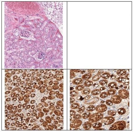 Immunohistochemistry (Formalin/PFA-fixed paraffin-embedded sections) - Anti-ALDH1L1 antibody (ab235197)
Immunohistochemical analysis of mouse kidney tissue labeling ALDH1L1 with ab235197 at 1/200 dilution, followed by a Donkey Anti-Goat IgG (HRP) secondary antibody for 20 minutes at room temperature. Images are shown at 20x (lower left) and 40x (lower right) magnification. Counter stained with Hematoxylin. Upper left panel, H&E staining.
Pathologist analysis: ALDH1L1 showed selective staining of renal tubular epithelial cells in mouse kidney. The specificity of the staining is cytoplasmic and membranous in tubules not in glomeruli which is consistent with ALDH1L1 staining in the Human Protein Atlas and as such, is suitable for IHC at a concentration of 1:200.
-
 Immunohistochemistry (Formalin/PFA-fixed paraffin-embedded sections) - Anti-ALDH1L1 antibody (ab235197)
Immunohistochemical analysis of mouse liver tissue labeling ALDH1L1 with ab235197 at 1/200 dilution, followed by a Donkey Anti-Goat IgG (HRP) secondary antibody for 20 minutes at room temperature. Images are shown at 20x (lower left) and 40x (lower right) magnification. Counter stained with Hematoxylin. Upper left panel, H&E staining.
Pathologist analysis: ALDH1L1 showed selective cytoplasmic staining in mouse liver of hepatocytes. The specificity of the staining is weak in bile duct cells and strong membranous and cytoplasmic in hepatocytes which is consistent with ALDH1L1 staining in the Human Protein Atlas and as such, is suitable for IHC at a concentration of 1:200.
-
 Western blot - Anti-ALDH1L1 antibody (ab235197)All lanes : Anti-ALDH1L1 antibody (ab235197) at 1/1000 dilution
Lane 1 : NIH/3T3 (mouse embryo fibroblast cell line) whole cell lysate
Lane 2 : Mouse liver lysate
Lane 3 : ALDH1L1 HEK293T overexpressed lysate
Secondary
All lanes : 605-703-125 GOAT IgG (H&L) Antibody Peroxidase Conjugated Pre-adsorbed at 1/40000 dilution
Predicted band size: 99 kDa
Exposure time: 1 secondALDH1L1 detection in NIH-3T3 by WB weak under general lysate conditions. Optimal detection of protein in 3T3 cells at G2/M enriched cells (not tested).
-
 Western blot - Anti-ALDH1L1 antibody (ab235197)Anti-ALDH1L1 antibody (ab235197) at 1/1000 dilution + NIH/3T3 (mouse embryo fibroblast cell line) whole cell lysate
Secondary
Goat polyclonal to ALDH1L1 (ab235197) at 1/40000 dilution (605-703-125 GOAT IgG (H&L) Antibody Peroxidase Conjugated Pre-adsorbed)
Predicted band size: 99 kDa
Observed band size: 80, 34 kDa why is the actual band size different from the predicted?
Exposure time: 35 secondsALDH1L1 detection in NIH-3T3 by WB weak under general lysate conditions. Optimal detection of protein in 3T3 cells at G2/M enriched cells (not tested).