Anti-AFG3L2 antibody (ab154990)
")
Key features and details
- Rabbit polyclonal to AFG3L2
- Suitable for: ICC/IF, WB, IHC-P
- Reacts with: Mouse, Rat
- Isotype: IgG
Overview
-
Product name
Anti-AFG3L2 antibody
See all AFG3L2 primary antibodies -
Description
Rabbit polyclonal to AFG3L2 -
Host species
Rabbit -
Tested applications
Suitable for: ICC/IF, WB, IHC-Pmore details -
Species reactivity
Reacts with: Mouse, Rat
Predicted to work with: Sheep, Rabbit, Cow, Cat, Chimpanzee, Macaque monkey, Gorilla, Orangutan
-
Immunogen
Synthetic peptide corresponding to Mouse AFG3L2 aa 50-150 conjugated to keyhole limpet haemocyanin.
Database link: Q8JZQ2 -
Positive control
- This antibody gave a positive signal in the following tissue lysates: Mouse Brain; Rat Brain; Mouse Hippocampus; Rat Hippocampus; Mouse Cerebellum. This antibody gave a positive result in IHC in the following FFPE tissue: Normal mouse brain. This antibody gave a positive result when used in the following methanol fixed cell lines: PC12
-
General notes
Reproducibility is key to advancing scientific discovery and accelerating scientists’ next breakthrough.
Abcam is leading the way with our range of recombinant antibodies, knockout-validated antibodies and knockout cell lines, all of which support improved reproducibility.
We are also planning to innovate the way in which we present recommended applications and species on our product datasheets, so that only applications & species that have been tested in our own labs, our suppliers or by selected trusted collaborators are covered by our Abpromise™ guarantee.
In preparation for this, we have started to update the applications & species that this product is Abpromise guaranteed for.
We are also updating the applications & species that this product has been “predicted to work with,” however this information is not covered by our Abpromise guarantee.
Applications & species from publications and Abreviews that have not been tested in our own labs or in those of our suppliers are not covered by the Abpromise guarantee.
Please check that this product meets your needs before purchasing. If you have any questions, special requirements or concerns, please send us an inquiry and/or contact our Support team ahead of purchase. Recommended alternatives for this product can be found below, as well as customer reviews and Q&As.
Properties
-
Form
Liquid -
Storage instructions
Shipped at 4°C. Store at +4°C short term (1-2 weeks). Upon delivery aliquot. Store at -20°C or -80°C. Avoid freeze / thaw cycle. -
Storage buffer
pH: 7.40
Preservative: 0.02% Sodium azide
Constituent: PBS
Batches of this product that have a concentration Concentration information loading...
Purity
Immunogen affinity purifiedClonality
PolyclonalIsotype
IgGResearch areas
Associated products
-
Compatible Secondaries
-
Isotype control
Applications
Our Abpromise guarantee covers the use of ab154990 in the following tested applications.
The application notes include recommended starting dilutions; optimal dilutions/concentrations should be determined by the end user.
Application Abreviews Notes ICC/IF Use a concentration of 5 µg/ml. WB Use a concentration of 1 µg/ml. Detects a band of approximately 89 kDa (predicted molecular weight: 89 kDa). Abcam recommends using milk as the blocking agent. Abcam welcomes customer feedback and would appreciate any comments regarding this product.
IHC-P Use a concentration of 5 µg/ml. Perform heat mediated antigen retrieval with citrate buffer pH 6 before commencing with IHC staining protocol. Target
-
Function
ATP-dependent protease which is essential for axonal development. -
Tissue specificity
Ubiquitous. Highly expressed in the cerebellar Purkinje cells. -
Involvement in disease
Defects in AFG3L2 are the cause of spinocerebellar ataxia type 28 (SCA28) [MIM:610246]. It is a clinically and genetically heterogeneous group of cerebellar disorders. Patients show progressive incoordination of gait and often poor coordination of hands, speech and eye movements, due to degeneration of the cerebellum with variable involvement of the brainstem and spinal cord. SCA28 is an autosomal dominant cerebellar ataxia (ADCA) with a slow progressive course and no evidence of sensory involvement or cognitive impairment.
Defects in AFG3L2 are the cause of spastic ataxia autosomal recessive type 5 (SPAX5) [MIM:614487]. A neurodegenerative disorder characterized by early onset spasticity, peripheral neuropathy, ptosis, oculomotor apraxia, dystonia, cerebellar atrophy, and progressive myoclonic epilepsy. -
Sequence similarities
In the N-terminal section; belongs to the AAA ATPase family.
In the C-terminal section; belongs to the peptidase M41 family. -
Cellular localization
Mitochondrion membrane. - Information by UniProt
-
Database links
- Entrez Gene: 515757 Cow
- Entrez Gene: 69597 Mouse
- Entrez Gene: 307350 Rat
- SwissProt: Q2KJI7 Cow
- SwissProt: Q8JZQ2 Mouse
- Unigene: 426052 Mouse
- Unigene: 8386 Rat
-
Alternative names
- AFG3 (ATPase family gene 3, yeast) like 2 antibody
- AFG3 ATPase family gene 3 like 2 (yeast) antibody
- AFG3 ATPase family gene 3 like 2 antibody
see all
Images
-
Western blot - Anti-AFG3L2 antibody (ab154990)All lanes : Anti-AFG3L2 antibody (ab154990) at 1 µg/ml (Milk blocking 3%)
Lane 1 : Brain (Mouse) Tissue Lysate
Lane 2 : Brain (Rat) Tissue Lysate
Lane 3 : Cerebellum Rat Tissue Lysate
Lane 4 : Mouse Hippocampus Tissue Lysate
Lane 5 : Rat Hippocampus Tissue Lysate
Lysates/proteins at 25 µg per lane.
Secondary
All lanes : Goat Anti-Rabbit IgG H&L (HRP) (ab97051) at 1/50000 dilution
Developed using the ECL technique.
Performed under reducing conditions.
Predicted band size: 89 kDa
Observed band size: 89 kDa
Additional bands at: 68 kDa (possible non-specific binding)
Exposure time: 2 minutesThis blot was produced using a 4-12% Bis-tris gel under the MOPS buffer system. The gel was run at 200V for 50 minutes before being transferred onto a Nitrocellulose membrane at 30V for 70 minutes. The membrane was then blocked for an hour using 3% Milk before being incubated with ab154990 overnight at 4°C. Antibody binding was detected using an anti-rabbit antibody conjugated to HRP, and visualised using ECL development solution ab133406.
-
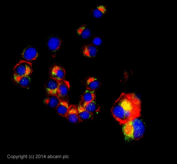 Immunocytochemistry/ Immunofluorescence - Anti-AFG3L2 antibody (ab154990)
ICC/IF image of ab154990 stained PC12 cells. The cells were 100% methanol fixed (5 min) and then incubated in 1%BSA / 10% normal goat serum / 0.3M glycine in 0.1% PBS-Tween for 1h to permeabilise the cells and block non-specific protein-protein interactions. The cells were then incubated with the antibody ab154990 at 5µg/ml overnight at +4°C. The secondary antibody (pseudo-colored green) was Alexa Fluor® 488 goat anti- rabbit (ab150081) IgG (H+L) preadsorbed, used at a 1/1000 dilution for 1h. Alexa Fluor® 594 WGA was used to label plasma membranes (pseudo-colored red) at a 1/200 dilution for 1h at room temperature. DAPI was used to stain the cell nuclei (pseudo-colored blue) at a concentration of 1.43µM for 1hour at room temperature.
-
 Immunohistochemistry (Formalin/PFA-fixed paraffin-embedded sections) - Anti-AFG3L2 antibody (ab154990)
IHC image of AFG3L2 staining in Normal mouse brain formalin fixed paraffin embedded tissue section*, performed on a Leica Bond™ system using the standard protocol B. The section was pre-treated using heat mediated antigen retrieval with sodium citrate buffer (pH6, epitope retrieval solution 1) for 20 mins. The section was then incubated with ab154990, 5µg/ml, for 15 mins at room temperature. A Goat anti-Rabbit biotinylated secondary antibody was used to detect the primary, and visualized using an HRP conjugated ABC system. DAB was used as the chromogen. The section was then counterstained with haematoxylin and mounted with DPX.
For other IHC staining systems (automated and non-automated) customers should optimize variable parameters such as antigen retrieval conditions, primary antibody concentration and antibody incubation times.
Protocols
Datasheets and documents
References (0)
ab154990 has not yet been referenced specifically in any publications.
Images
-
Western blot - Anti-AFG3L2 antibody (ab154990)All lanes : Anti-AFG3L2 antibody (ab154990) at 1 µg/ml (Milk blocking 3%)
Lane 1 : Brain (Mouse) Tissue Lysate
Lane 2 : Brain (Rat) Tissue Lysate
Lane 3 : Cerebellum Rat Tissue Lysate
Lane 4 : Mouse Hippocampus Tissue Lysate
Lane 5 : Rat Hippocampus Tissue Lysate
Lysates/proteins at 25 µg per lane.
Secondary
All lanes : Goat Anti-Rabbit IgG H&L (HRP) (ab97051) at 1/50000 dilution
Developed using the ECL technique.
Performed under reducing conditions.
Predicted band size: 89 kDa
Observed band size: 89 kDa
Additional bands at: 68 kDa (possible non-specific binding)
Exposure time: 2 minutesThis blot was produced using a 4-12% Bis-tris gel under the MOPS buffer system. The gel was run at 200V for 50 minutes before being transferred onto a Nitrocellulose membrane at 30V for 70 minutes. The membrane was then blocked for an hour using 3% Milk before being incubated with ab154990 overnight at 4°C. Antibody binding was detected using an anti-rabbit antibody conjugated to HRP, and visualised using ECL development solution ab133406.
-
Immunocytochemistry/ Immunofluorescence - Anti-AFG3L2 antibody (ab154990)
ICC/IF image of ab154990 stained PC12 cells. The cells were 100% methanol fixed (5 min) and then incubated in 1%BSA / 10% normal goat serum / 0.3M glycine in 0.1% PBS-Tween for 1h to permeabilise the cells and block non-specific protein-protein interactions. The cells were then incubated with the antibody ab154990 at 5µg/ml overnight at +4°C. The secondary antibody (pseudo-colored green) was Alexa Fluor® 488 goat anti- rabbit (ab150081) IgG (H+L) preadsorbed, used at a 1/1000 dilution for 1h. Alexa Fluor® 594 WGA was used to label plasma membranes (pseudo-colored red) at a 1/200 dilution for 1h at room temperature. DAPI was used to stain the cell nuclei (pseudo-colored blue) at a concentration of 1.43µM for 1hour at room temperature.
-
Immunohistochemistry (Formalin/PFA-fixed paraffin-embedded sections) - Anti-AFG3L2 antibody (ab154990)
IHC image of AFG3L2 staining in Normal mouse brain formalin fixed paraffin embedded tissue section*, performed on a Leica Bond™ system using the standard protocol B. The section was pre-treated using heat mediated antigen retrieval with sodium citrate buffer (pH6, epitope retrieval solution 1) for 20 mins. The section was then incubated with ab154990, 5µg/ml, for 15 mins at room temperature. A Goat anti-Rabbit biotinylated secondary antibody was used to detect the primary, and visualized using an HRP conjugated ABC system. DAB was used as the chromogen. The section was then counterstained with haematoxylin and mounted with DPX.
For other IHC staining systems (automated and non-automated) customers should optimize variable parameters such as antigen retrieval conditions, primary antibody concentration and antibody incubation times.


