Anti-53BP1 antibody (ab172580)
")
Key features and details
- Rabbit polyclonal to 53BP1
- Suitable for: WB, ICC/IF, IP, Flow Cyt, IHC-P
- Reacts with: Mouse, Rat, Human
- Isotype: IgG
Overview
-
Product name
Anti-53BP1 antibody
See all 53BP1 primary antibodies -
Description
Rabbit polyclonal to 53BP1 -
Host species
Rabbit -
Tested Applications & Species
See all applications and species dataApplication Species Flow Cyt HumanICC/IF HumanIHC-P HumanIP HumanWB Human
-
Immunogen
Synthetic peptide corresponding to Human 53BP1 aa 1400-1700.
-
General notes
The Life Science industry has been in the grips of a reproducibility crisis for a number of years. Abcam is leading the way in addressing the problem with our range of recombinant monoclonal antibodies and knockout edited cell lines for gold-standard validation.
One factor contributing to the crisis is the use of antibodies that are not suitable. This can lead to misleading results and the use of incorrect data informing project assumptions and direction. To help address this challenge, we have introduced an application and species grid on our primary antibody datasheets to make it easy to simplify identification of the right antibody for your needs.
Learn more here.
Properties
-
Form
Liquid -
Storage instructions
Shipped at 4°C. Store at -20°C. -
Storage buffer
Constituent: Water -
 Concentration information loading...
 -
Purity
Tissue culture supernatant -
Clonality
Polyclonal -
Isotype
IgG -
Research areas
Images
-
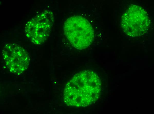
Immunocytochemistry/ Immunofluorescence - Anti-53BP1 antibody (ab172580)
Immunocytochemistry/Immunofluorescence analysis of U2OS cells labelling 53BP1 with ab172580 at 1:1000. Cells were fixed with PFA and permeabilized with methanol for 2 minutes. Cells were incubated with the primary anitbody overnight at room temperature. Top - 53BP1, Middle - DAPI, Bottom - Merge.
-
 Immunocytochemistry/ Immunofluorescence - Anti-53BP1 antibody (ab172580)
Immunocytochemistry/Immunofluorescence analysis of U2OS cells labelling 53BP1 with ab172580 at 1:1000. Cells were fixed with PFA and permeabilized with methanol for 2 minutes. Cells were incubated with the primary anitbody overnight at room temperature.
-
 Flow Cytometry - Anti-53BP1 antibody (ab172580)
Flow Cytometry analysis of HeLa cells labelling 53BP1 with ab172580 at 1:800. Cells were fixed with 1% PFA. Cells were incuabted with the primary antibody overnight at 4°C.
-
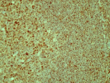 Immunohistochemistry (Formalin/PFA-fixed paraffin-embedded sections) - Anti-53BP1 antibody (ab172580)
Immunohistochemistry (formalin/PFA-fixed paraffin-embedded) analysis of Human linfoid tissue labelling 53BP1 with ab172580 at 1:1000. Antigen retrieval was by microwave boiling for 7 minutes in 10mM TrisHCl, pH9.
-
 Immunoprecipitation - Anti-53BP1 antibody (ab172580)
Immunoprecipitation analysis of U2OS cells labelling 53BP1 with ab172580. Protein A beads were crosslinked to the serum.
-
 Western blot - Anti-53BP1 antibody (ab172580)All lanes : Anti-53BP1 antibody (ab172580)
Lane 1 : U2OS (Control si)
Lane 2 : U2OS (53BP1 si)
Lane 3 : NIH 3T3 cell lysate at 40 µg
Lane 4 : PC12 cell lysate at 40 µg
Predicted band size: 214 kDa


