AKT total + Phospho S473 FLOW Kit (ab126580)
")
Key features and details
- Assay type: Direct
- Detection method: Fluorescent
- Platform: Flow cytometer
- Sample type: Adherent cells, Suspension cells
Overview
-
Product name
AKT total + Phospho S473 FLOW Kit -
Detection method
Fluorescent -
Sample type
Adherent cells, Suspension cells -
Assay type
Direct -
Species reactivity
Reacts with: Mouse, Human -
Product overview
Akt is a serine, threonine protein kinase critical in cellular metabolism, glucose uptake, protein synthesis, cell proliferation, growth, apoptosis, survival, angiogenesis, migration and invasion. It acts downstream of the phosphatidylinositol 3 kinase (PI3K) and it mediates the effects of several growth factors such as platelet-derived growth factor, epidermal growth factor and insulin growth factor. It is activated by phosphorylation on Ser-473, Thr-308 and Tyr-474 and when active it phosphorylates transcription factors (FOXO1), kinases (GSK-3, Raf-1, ASK, Chk1) and other signaling proteins (Bad, MDM2). There are three Akt isoforms (Akt1, Akt2 and Akt3) which share 80% sequence identity also known as PKBα, PKBβ and PKBγ. Akt has been shown to have a role in metabolism, apoptosis and proliferation and therefore it has been proposed to be the candidate “Warburg Kinase”.
-
Platform
Flow cytometer
Properties
-
Storage instructions
Store at +4°C. Please refer to protocols. -
Components 50 tests 10X Phosphate Buffered Saline 1 x 100ml 400X Tween-20 1 x 4ml 50X Antibody cocktail 1 x 100µl Blocking Solution 1 x 10ml -
Research areas
-
Function
IGF-1 leads to the activation of AKT3, which may play a role in regulating cell survival. Capable of phosphorylating several known proteins. Truncated isoform 2/PKB gamma 1 without the second serine phosphorylation site could still be stimulated but to a lesser extent. -
Tissue specificity
In adult tissues, it is highly expressed in brain, lung and kidney, but weakly in heart, testis and liver. In fetal tissues, it is highly expressed in heart, liver and brain and not at all in kidney. -
Sequence similarities
Belongs to the protein kinase superfamily. AGC Ser/Thr protein kinase family. RAC subfamily.
Contains 1 AGC-kinase C-terminal domain.
Contains 1 PH domain.
Contains 1 protein kinase domain. -
Domain
Binding of the PH domain to the phosphatidylinositol 3-kinase alpha (PI(3)K) results in its targeting to the plasma membrane. -
Post-translational
modificationsPhosphorylation on Thr-305 and Ser-472 is required for full activity (By similarity). Phosphorylated upon DNA damage, probably by ATM or ATR.
Ubiquitinated. When fully phosphorylated and translocated into the nucleus, undergoes 'Lys-48'-polyubiquitination catalyzed by TTC3, leading to its degradation by the proteasome. -
Cellular localization
Cytoplasm. Membrane. Membrane-associated after cell stimulation leading to its translocation. - Information by UniProt
-
Alternative names
- Akt3
- AKT3_HUMAN
- PKB gamma
see all -
Database links
- Entrez Gene: 208 Human
- Entrez Gene: 207 Human
- Entrez Gene: 10000 Human
- Entrez Gene: 23797 Mouse
- SwissProt: P31751 Human
- SwissProt: Q9Y243 Human
- SwissProt: Q9WUA6 Mouse
- Unigene: 498292 Human
see all
Images
-
Flow Cytometry - AKT total (s473) FLOW Kit (ab126580)Figure 1. Antibody specificity demonstrated by Flow cytometry. Non induced (red) and PDGF induced (blue) NIH3T3 were targeted with the antibody cocktail against Akt1. Background fluorescence (black) was determined with a no-primary antibody control. After background subtraction, the PDGF induced cell line shows 1.2 increase in the levels of Akt1 protein.
-
 Flow Cytometry - AKT total (s473) FLOW Kit (ab126580)Figure 2. Antibody specificity demonstrated by Flow cytometry. Non induced (red) and PDGF induced (blue) NIH3T3 were targeted with the antibody cocktail against Akt1 (phospho S473). Background fluorescence (black) was determined with a no-primary antibody control. After background subtraction, the PDGF induced cell line shows a 7 fold increase in the levels of phosphorylated Akt1.
 -
 Flow Cytometry - AKT total (s473) FLOW Kit (ab126580)Figure 3. Antibody specificity demonstrated by Flow cytometry. Non induced (vehicle) and PDGF induced NIH3T3 were targeted with the antibody cocktail against Akt1. Background fluorescence was determined with a no-primary antibody control. After background subtraction, the PDGF induced cell line shows 1.2 increase in the levels of Akt1 protein.
 -
 Flow Cytometry - AKT total (s473) FLOW Kit (ab126580)Figure 4. Antibody specificity demonstrated by Flow cytometry. Non induced (vehicle) and PDGF induced NIH3T3 were targeted with the antibody cocktail against Akt1 (phospho S473). Background fluorescence was determined with a no-primary antibody control. After background subtraction, the PDGF induced cell line shows a 7 fold increase in the levels of phosphorylated Akt1.
 -
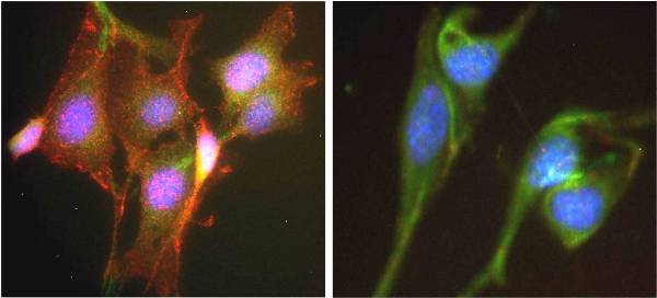 ICC AKT Total and pSer473Antibody specificity demonstrated by immunocytochemistry. ICC was carried out on NIH3T3 cells treated with PDGF (Left) or vehicle (right) with anti-Akt1 phosphoS473 and anti-Akt1 and all buffer reagents as supplied in this kit. Labeling was carried out with a polyclonal antibody GAR-594 and GAM-488 respectively. The PDGF induced cells (left) show a significant induction of Akt phosphorylation at residue S473 (observed in the 594 channel) in comparison to the non-induced control (right).
 -
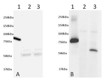 Western blot - AKT total (s473) FLOW Kit (ab126580)Figure 5. Validation of antibodies by Western Blot. Western blot was run on a 10-20% gradient acrylamide gel. Samples were loaded as follows from left to right: (1) 50ng of Human recombinant AKT1 protein (tagged) (ab62279), (2) 25ug of non-induced NIH3T3 cell extract and (3) 25ug of PDGF induced NIH3T3 cell extract. Membrane Blocking was carried out with 5% Milk+50mM Tris+0.05% Tween-20 pH 7.4, primary antibodies (ab54752 at 5ug/mL left and ab81283 at 1:5000 right) were incubated overnight in 5% BSA+50mM+0.05% Tween-20 pH 7.4 and secondary antibodies were incubated for 2 hours in 5% Milk+50mM Tris+0.05% Tween-20 pH 7.4.


