Organelle Detection Western Blot Cocktail (ab133989)
Key features and details
- Assay type: Quantitative
- Sample type: Adherent cells, Cell culture extracts, Cell Lysate, Nuclear Extracts, Suspension cells, Tissue Extracts, Tissue Homogenate
Overview
-
Product name
Organelle Detection Western Blot Cocktail -
Sample type
Cell culture extracts, Adherent cells, Suspension cells, Tissue Extracts, Cell Lysate, Tissue Homogenate, Nuclear Extracts -
Assay type
Quantitative -
Species reactivity
Reacts with: Mouse, Rat, Human -
Product overview
ab133989 contains 4 mAbs each targeting a specific organelle marker. The presence of plasma membrane is determined by Anti-Sodium Potassium ATPase antibody; mitochondrion by Anti-ATP5A antibody; cytosol by Anti-GAPDH; and nucleus by Anti-Histone H3 (di methyl K9). This cocktail is suitable for determining the purity of organelle isolates prior to further characterization.
This product is particularly valuable to researchers working in organelle proteomics. Mass spectrometry is frequently used in this field to determine the protein content of targeted organelle isolates. These isolates are obtained using differential centrifugation, density gradient fractionation, biochemical enrichment, or affinity purification. Unfortunately, the various methods of purification available for organelle isolation are imperfect and leave behind contaminants from undesired regions of the cell. These contaminants are inevitable, but being aware of which contaminants are present is crucial for analysis of mass spectrometry results. The high sensitivity and species cross reactivity of the antibodies in this cocktail will quickly and easily reveal impurities caused by imperfect sample preparation.
-
Tested applications
Suitable for: WBmore details
Images
-
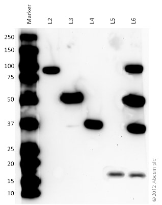
All blocking and antibody incubation steps were done in 5% milk, 20 mM Tris-HCl, 0.1% TWEEN-20.
Lane 2-6 : Mouse heart homogenate Whole Tissue Lysate 10 µg
Primary antibody:
Lane 2 : Anti-Sodium Potassium ATPase antibody – Plasma Membrane Marker
Lane 3 : Anti-ATP5A antibody – Mitochondrial Marker
Lane 4 : Anti-GAPDH antibody – Cytosolic Marker
Lane 5 : Anti-Histone H3 (di methyl K9) antibody – Nuclear Marker
Lane 6 : Assembled Organelle Detection Cocktail
Secondary: ab131368 at 1/1000 dilution.
Predicted Sodium Potassium ATPase band size : 113 kDa
Observed band size : 85 kDa
Predicted ATP5A band size : 60 kDa
Observed ATP5A band size : 52 kDa
Predicted sample band size : 36 kDa
Observed band size : 36 kDa
Predicted sample band size : 15.5 kDa
Observed band size : 17 kDa -
All blocking and antibody incubation steps were done in 5% milk, 20 mM Tris-HCl, 0.1% TWEEN-20.
All lanes :
Anti-Sodium Potassium ATPase antibody – Plasma Membrane Marker
Anti-ATP5A antibody – Mitochondrial Marker
Anti-GAPDH antibody – Cytosolic Marker
Anti-Histone H3 (di methyl K9) antibody – Nuclear Marker
Lane 1 : Marker
Lane 2 : Human heart homogenate Whole Tissue Lysate - 20 µg
Lane 3 : HeLa Whole Cell Lysate - 20 µg
Lane 4 : Mouse heart homogenate Whole Tissue Lysate - 20 µg
Lane 5 : NIH-3T3 Whole Cell Lysate - 20 µg
Lane 6 : Rat heart homogenate Whole Tissue Lysate - 20 µg
Lane 7 : H9C2 Whole Cell Lysate - 20 µg
Secondary: ab131368 at 1/1000 dilution. -
HeLa cell lysate was prepared using the Cell Fractionation Kit ab109719. All blocking and antibody incubation steps were done in 5% milk, 20 mM Tris-HCl, 0.1% TWEEN-20.
All lanes :
Anti-Sodium Potassium ATPase antibody – Plasma Membrane Marker
Anti-ATP5A antibody – Mitochondrial Marker
Anti-GAPDH antibody – Cytosolic Marker
Anti-Histone H3 (di methyl K9) antibody – Nuclear Marker
Lane 1 : Marker
Lane 2 : HeLa Whole Cell Lysate
Lane 3 : HeLa Cytosolic Fraction Lysate
Lane 4 : HeLa Mitochondrial Fraction Lysate
Lane 5 : HeLa Nuclear Fraction Lysate
Secondary
Goat polyclonal to Mouse IgG – H&L – Pre-Adsorbed (HRP) at 1/10000.


