Normal Goat Serum (ab7481)
Overview
-
Product name
Normal Goat Serum
See all Goat Serum reagents -
Host species
Goat -
Tested applications
Suitable for: Blocking, IHC-Fr, ICC/IF, IHC-Pmore details -
General notes
Normal goat serum ab7481 is used extensively for the blocking of non-specific antibody binding in tissue and cell staining, and in other applications of antibodies.
The goat serum blocks the binding of Fc receptors in the sample to the primary and secondary antibodies used in the experiment, and also blocks non-specific binding of the antibodies to the sample.
Typically the serum used for blocking is from a different species than the species in which the primary antibody was raised. Often the blocking serum is from the species in which the secondary antibody was raised.
Serum can be used directly for blocking, or as a constituent of a blocking buffer.
Strain: Mixed breed and sex.
Raised in: Goat
Purity: Whole antiserum
This product is for research use only and not intended for diagnostic or therapeutic use of any kind.
Properties
-
Form
Liquid -
Storage instructions
Shipped on Dry Ice. Upon delivery aliquot. Store at -20°C or -80°C. Avoid freeze / thaw cycle. -
Storage buffer
Constituent: Whole serum -
 Concentration information loading...
 -
Purity
Whole antiserum -
Reagent notes
Strain: Mixed breed and sex. -
Research areas
Images
-
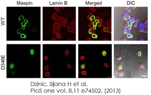
 Functional Studies - Normal Goat Serum (ab7481) Dzinic, Sijana H et al., PloS one vol. 8,11 e74502., Fig 2, doi:10.1371/journal.pone.0074502
Cells grown in 8-well chamber slides to 70% confluence were fixed with 4% paraformaldehyde (15 min at room temperature (RT)), and permeabilized with 100% ice cold methanol (10 min at 20°C). The slides were incubated with 10% normal goat serum (ab7481) in PBS for 1 hr, and incubated with anti-maspin (1:100) antibody alone or in a combination with either anti-lamin B (1:50), anti-HDAC1 (1:50) or anti-GRP78 (1:50) at 4°C overnight. Cells were washed and incubated for 2 hrs at room temperature (RT) with Alexa Fluor 488 (1:500) alone or in combination with Alexa Fluor 594 (1:500). The nuclei were counterstained with DAPI.
DU145 cells infected with adenovirus expressing either maspinWT or maspinD346E Confocal immunofluorescence imaging of maspin (green) and nuclear envelope marker lamin B (red) in DU145 cells after infection. -
 Functional Studies - Normal Goat Serum (ab7481) Witasp, Anna et al., PloS one vol. 8,5 e63493., Fig 3, doi:10.1371/journal.pone.0063493Slides were pretreated with Hydrogen Peroxide Block followed by 15% normal goat serum (Abcam, Cambridge, UK) for 1 hour. The primary antibody, anti-PTX3, N-terminal antibody produced in rabbit was diluted 1:300 in PBS with 2.5% goat serum (ab7481), applied to slides and incubated for 3 hours at 4°C. The primary antibody was omitted in the negative controls. The PTX3 binding was revealed using a universal secondary antibody polymer formulation conjugated to horseradish peroxidase (HRP). The HRP activity was subsequently visualized with diaminobenzidine (DAB) substrate/chromogen and counterstaining with hematoxylin was performed.


