Neuraminidase Assay Kit (Fluorometric - Blue) (ab138888)
Key features and details
- Assay type: Enzyme activity
- Detection method: Fluorescent
- Platform: Microplate reader
- Sample type: Adherent cells, Cell culture extracts, Cell culture supernatant, Suspension cells, Tissue Culture Media
- Sensitivity: 0.3 mU/ml
Overview
-
Product name
Neuraminidase Assay Kit (Fluorometric - Blue)
See all Neuraminidase kits -
Detection method
Fluorescent -
Sample type
Cell culture supernatant, Cell culture extracts, Adherent cells, Suspension cells, Tissue Culture Media -
Assay type
Enzyme activity -
Sensitivity
= 0.3 mU/ml -
Species reactivity
Reacts with: Mammals, Other species -
Product overview
Neuraminidase Assay Kit (Fluorometric - Blue) (ab138888) provides a sensitive and robust fluorometric assay to detect neuraminidase that exists either in cells or biological samples. The non-fluorescent neuraminidase substrate becomes strongly fluorescent upon neuraminidase cleavage. The kit can detect as little as 0.3 mU/mL neuraminidase in a 100 µL assay volume. The assay can be performed in a convenient 96-well or 384-well microtiter-plate format and easily adapted to automation without a separation step. The signal can be easily read by a fluorescence microplate reader at Ex/Em = ~320/~450 nm.
-
Notes
Neuraminidases, also called sialidases, are glycoside hydrolase enzymes that catalyze the hydrolysis of terminal sialic acid residues and neuraminic acids. The most commonly known neuraminidase is the viral neuraminidase. The cleavage of linkage between sialic acid and adjacent sugar residue permits the transport of the virus through mucin and destroys the haemagglutinin receptor on the host cell, thus allowing elution of progeny virus particles from infected cells. Neuraminidase promotes influenza virus release from infected cells and facilitates virus spread within the respiratory tract. Thus, it is an important target for influenza drug development. The detection of neuraminidase and screening its inhibitors is one of the essential tasks for investigating biological processes and prevention of influenza infection.
-
Platform
Microplate reader
Properties
-
Storage instructions
Store at -20°C. Please refer to protocols. -
Components 200 tests Assay Buffer 1 x 20ml Neuraminidase Standard 1 x 0.1 unit NeuroBlue Indicator 1 vial -
Research areas
-
Function
Catalyzes the removal of sialic acid (N-acetylneuramic acid) moities from glycoproteins and glycolipids. To be active, it is strictly dependent on its presence in the multienzyme complex. Appears to have a preference for alpha 2-3 and alpha 2-6 sialyl linkage. -
Tissue specificity
Highly expressed in pancreas, followed by skeletal muscle, kidney, placenta, heart, lung and liver. Weakly expressed in brain. -
Involvement in disease
Defects in NEU1 are the cause of sialidosis (SIALIDOSIS) [MIM:256550]. It is a lysosomal storage disease occurring as two types with various manifestations. Type 1 sialidosis (cherry red spot-myoclonus syndrome or normosomatic type) is late-onset and it is characterized by the formation of cherry red macular spots in childhood, progressive debilitating myoclonus, insiduous visual loss and rarely ataxia. The diagnosis can be confirmed by the screening of the urine for sialyloligosaccharides. Type 2 sialidosis (also known as dysmorphic type) occurs as several variants of increasing severity with earlier age of onset. It is characterized by the presence of abnormal somatic features including coarse facies and dysostosis multiplex, vertebral deformities, mental retardation, cherry-red spot/myoclonus, sialuria, cytoplasmic vacuolation of peripheral lymphocytes, bone marrow cells and conjunctival epithelial cells. -
Sequence similarities
Belongs to the glycosyl hydrolase 33 family.
Contains 4 BNR repeats. -
Domain
A C-terminal internalization signal (YGTL) appears to allow the targeting of plasma membrane proteins to endosomes. -
Post-translational
modificationsN-glycosylated.
Phosphorylation of tyrosine within the internalization signal results in inhibition of sialidase internalization and blockage on the plasma membrane. -
Cellular localization
Lysosome membrane. Lysosome lumen. Cell membrane. Cytoplasmic vesicle. Localized not only on the inner side of the lysosomal membrane and in the lysosomal lumen, but also on the plasma membrane and in intracellular vesicles. - Information by UniProt
-
Alternative names
- Acetylneuraminyl hydrolase
- exo-alpha-sialidase
- G9 sialidase
see all
Images
-
Neuraminidase Activity measured in mouse tissue lysates.
Protein concentration for samples varied from 7 mg/mL to 30 mg/mL. Samples were diluted 10-30 fold.
-
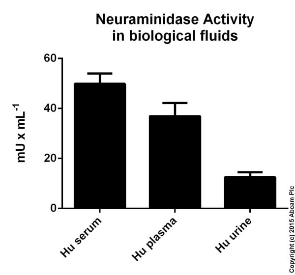
Neuraminidase Activity measured in biological fluids. The data are averages of values obtained from several sample dilutions, assayed in duplicate. Serum samples were diluted 1/10 and 1/30, plasma samples were diluted 1/10, and urine samples were undiluted and diluted 1/3 and 1/9.
-
Neuraminidase dose response was measured in a 96-well black plate with Neuraminidase Assay Kit (Fluorometric –Blue) using a Germini fluorescence microplate reader (Molecular Devices). As low as 0.3 mU/mL of neuraminidase can be detected with 1 hour incubation time in 37°C, 5% CO2 incubator.


