Mouse IL-1ra ELISA Kit (ab113348)
Key features and details
- Sensitivity: 2 pg/ml
- Range: 2.74 pg/ml - 2000 pg/ml
- Sample type: Cell culture supernatant, Plasma, Serum
- Detection method: Colorimetric
- Assay type: Sandwich (quantitative)
- Reacts with: Mouse
Properties
-
Storage instructions
Store at -20°C. Please refer to protocols. -
Components 1 x 96 tests 20X Wash Buffer Concentrate 1 x 25ml 300X HRP-Streptavidin Concentrate 1 x 200µl 5X Assay Diluent B 1 x 15ml 5X Assay Diluent D 1 x 15ml Biotinylated anti-Mouse IL-1ra 2 vials IL-1ra Microplate (12 x 8 wells) 1 unit Recombinant Mouse IL-1ra Standard (lyophilized) 2 vials Stop Solution 1 x 8ml TMB One-Step Substrate Reagent 1 x 12ml -
Research areas
-
Function
Inhibits the activity of interleukin-1 by binding to receptor IL1R1 and preventing its association with the coreceptor IL1RAP for signaling. Has no interleukin-1 like activity. Binds functional interleukin-1 receptor IL1R1 with greater affinity than decoy receptor IL1R2; however, the physiological relevance of the latter association is unsure. -
Tissue specificity
The intracellular form of IL1RN is predominantly expressed in epithelial cells. -
Involvement in disease
Microvascular complications of diabetes 4
Interleukin 1 receptor antagonist deficiency -
Cellular localization
Cytoplasm and Secreted. - Information by UniProt
-
Alternative names
- DIRA
- F630041P17Rik
- ICIL 1RA
see all -
Database links
- Entrez Gene: 3557 Human
- Entrez Gene: 16181 Mouse
- Entrez Gene: 60582 Rat
- Omim: 147679 Human
- SwissProt: P18510 Human
- SwissProt: P25085 Mouse
- SwissProt: P25086 Rat
- Unigene: 81134 Human
see all
Images
-
Standard curve of msIL-1Ra in different diluents with background signal subtracted (duplicates; +/- SD).
-
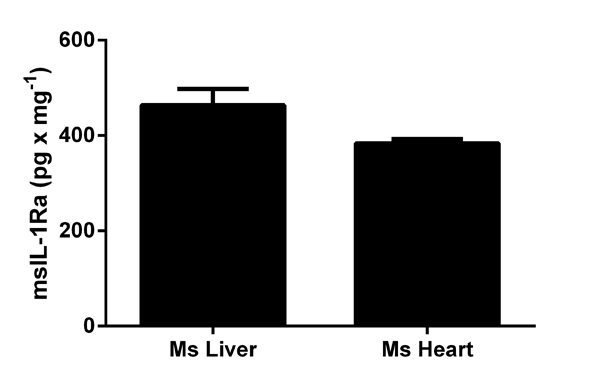
IL-1Ra measured in mouse tissue lysates (expressed as per mg of extracted protein), with background signal subtracted (duplicates; +/- SD).
-
msIL-1Ra measured in biological fluids, background signal subtracted (duplicates +/- SD).
-
IL-1Ra detected in supernatants from RAW 246.7 control cells (C) or cells stimulated for 24 hours with 50 ng x mL-1 of PMA (ab120297) (P), or 24 hours with PMA and 1 ug x mL-1 of LPS (Sigma) (P+L) for the last 6 hours. Results shown after background signal was subtracted (duplicates +/- SD).
-
Representative Standard Curve using ab113348
-
Representative Standard Curve using ab113348


