Mouse IL-1a ELISA Kit (ab199076)
Key features and details
- One-wash 90 minute protocol
- Sensitivity: 0.62 pg/ml
- Range: 4.69 pg/ml - 300 pg/ml
- Sample type: Cell culture supernatant, Cit plasma, Serum
- Detection method: Colorimetric
- Assay type: Sandwich (quantitative)
- Reacts with: Mouse
Overview
-
Product name
Mouse IL-1a ELISA Kit
See all IL-1 alpha kits -
Detection method
Colorimetric -
Precision
Intra-assay Sample n Mean SD CV% Overall 8 2.31% Inter-assay Sample n Mean SD CV% Overall 3 4.78% -
Sample type
Cell culture supernatant, Serum, Cit plasma -
Assay type
Sandwich (quantitative) -
Sensitivity
0.62 pg/ml -
Range
4.69 pg/ml - 300 pg/ml -
Recovery
Sample specific recovery Sample type Average % Range Cell culture supernatant 88.8 87.4% - 90.3% Serum 101.2 98.5% - 103.5% Cell culture media 94.4 93.4% - 96.1% Cit plasma 97.6 94.1% - 101.6% -
Assay time
1h 30m -
Assay duration
One step assay -
Species reactivity
Reacts with: Mouse -
Product overview
Mouse IL-1a ELISA Kit (ab199076) is a single-wash 90 min sandwich ELISA designed for the quantitative measurement of IL-1a protein in cell culture supernatant, cit plasma, and serum. It uses our proprietary SimpleStep ELISA® technology. Quantitate Mouse IL-1a with 0.62 pg/ml sensitivity.
SimpleStep ELISA® technology employs capture antibodies conjugated to an affinity tag that is recognized by the monoclonal antibody used to coat our SimpleStep ELISA® plates. This approach to sandwich ELISA allows the formation of the antibody-analyte sandwich complex in a single step, significantly reducing assay time. See the SimpleStep ELISA® protocol summary in the image section for further details. Our SimpleStep ELISA® technology provides several benefits:
- Single-wash protocol reduces assay time to 90 minutes or less
- High sensitivity, specificity and reproducibility from superior antibodies
- Fully validated in biological samples
- 96-wells plate breakable into 12 x 8 wells stripsA 384-well SimpleStep ELISA® microplate (ab203359) is available to use as an alternative to the 96-well microplate provided with SimpleStep ELISA® kits.
-
Notes
Interleukin 1 (IL-1) refers to two proteins encoded by two different genes (IL-1a and IL-1b), both of which share the same cell surface receptors. Mouse IL-1a is a pro-inflammatory cytokine that encodes a 270 aa residue called pro-IL-1a, which contains a nuclear localization sequence, an N-myristoylation site, and three potential N-linked glycosylation sites. Primarily, pro-IL-1a is localized to the cytosol after synthesis. However, some pro-IL-1a is secreted and is subsequently cleaved by extracellular proteases such as calpain to form the 17 kDa, 154 aa residue mature IL-1a. Importantly, both pro-IL-1a and mature IL-1a have been shown to bind the IL-1 receptor and are biologically active. This is different than pro-IL-1b and mature IL-1b, where pro-IL-1b is biologically inactive. Within the mature protein, rat IL-1a and human IL-1a share 79% and 56% aa sequence identity, respectively, with the mouse IL-1a protein.
IL-1a and IL-1b interact with three type I transmembrane Ig superfamily proteins: IL-1 receptor type I (IL-1 RI), IL-1 RII, and IL-1 receptor accessory protein (IL-1 RAcP). Both IL-1 RI and IL-1 RII can bind directly to IL-1a and IL-1b. IL-1 RAcP interacts with IL-1 RI in the presence of IL-1 to form a high-affinity receptor complex that is required for intracellular signal transduction. Interestingly, IL-1 RAcP will also bind IL-1 RII, but this creates a non-functional high-affinity receptor complex that does not transduce IL-1 signals. Thus, IL-1 RII can act to moderate the function of IL-1a and IL-1b.
IL-1a is typically expressed under normal conditions in skin keratinocytes and some epithelial cells, as well as in some cells in the central nervous system. Under pathological conditions, IL-1a is also strongly expressed by monocytes, tissue macrophages, dendritic cells, B lymphocytes and NK cells. Intracellular pro-IL-1a can also be localized to the nucleus where it plays a role as an intracellular regulator of human endothelial cell proliferation and migration. IL-1 possesses a wide variety of biological activities and plays a central role in mediating immune and inflammatory responses. Normal production of IL-1 is critical for hematopoiesis, osteoclast differentiation, and initiation of normal host responses to stresses such as injury and infection. Incorrect production of IL-1 has been shown to be associated with a variety of pathological conditions including sepsis, rheumatoid arthritis, inflammatory bowel disease, acute and chronic myelogenous leukemia, insulin-dependent diabetes mellitus, and atherosclerosis.
-
Platform
Microplate (12 x 8 well strips)
Properties
-
Storage instructions
Store at +4°C. Please refer to protocols. -
Components 1 x 96 tests 10X Mouse IL-1a (Interleukin-1 alpha) Capture Antibody 1 x 600µl 10X Mouse IL-1a (Interleukin-1 alpha) Detector Antibody 1 x 600µl 10X Wash Buffer PT (ab206977) 1 x 20ml Antibody Diluent CPI - HAMA Blocker (ab193969) 1 x 6ml Mouse IL-1a (Interleukin-1 alpha) Lyophilized Recombinant Protein (ab119727) 2 vials Plate Seals 1 unit Sample Diluent NS (ab193972) 1 x 50ml SimpleStep Pre-Coated 96-Well Microplate (ab206978) 1 unit Stop Solution 1 x 12ml TMB Development Solution 1 x 12ml -
Research areas
-
Relevance
IL-1 alpha is a member of the interleukin 1 cytokine family. It is a pleiotropic cytokine involved in various immune responses, inflammatory processes, and hematopoiesis. IL-1 alpha is produced by monocytes and macrophages as a proprotein, which is proteolytically processed and released in response to cell injury, and thus induces apoptosis. This gene and eight other interleukin 1 family genes form a cytokine gene cluster on chromosome 2. It has been suggested that the polymorphism of these genes is associated with rheumatoid arthritis and Alzheimer's disease. -
Cellular localization
Secreted -
Alternative names
- Hematopoietin 1
- IL 1
- IL 1 alpha
see all -
Database links
- Entrez Gene: 16175 Mouse
- SwissProt: P01582 Mouse
Images
-
SimpleStep ELISA technology allows the formation of the antibody-antigen complex in one single step, reducing assay time to 90 minutes. Add samples or standards and antibody mix to wells all at once, incubate, wash, and add your final substrate. See protocol for a detailed step-by-step guide.
-
Standard curve comparison between mouse IL-1a SimpleStep ELISA® kit and traditional ELISA kit from leading competitor. SimpleStep ELISA kit shows comparable sensitivity.
-
Background-subtracted data values (mean +/- SD) are graphed.
-
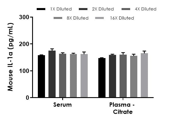
Recombinant IL-1a mouse was spiked into 50% mouse serum and 50% mouse plasma and diluted in a 2-fold dilution series in Sample Diluent NS. The concentrations of mouse IL-1a were measured in duplicate and interpolated from the mouse IL-1a standard curve and corrected for sample dilution. The interpolated dilution factor corrected values are graphed (mean +/- SD).
-
RAW264.7 cells were cultured in high glucose DMEM with 10% fetal calf serum or horse serum, 2 mM L-glutamine and 100 µg/mL Kanamycin. RAW264.7 cells were starved for 24 hours and treated in the presence and absence of 5 µg/mL of LPS. The concentrations of IL1-a were interpolated from the calibration curve and corrected for sample dilution (1:2). The mean IL-1a concentration in RAW264.7 media was determined to be 288 pg/mL in stimulated RAW 264.7 cells and undetectable in unstimulated RAW264.7 cells.
-
To learn more about the advantages of recombinant antibodies see here.


