Human MIP3a ELISA Kit (CCL20) (ab178015)
 (ab178015)")
Key features and details
- One-wash 90 minute protocol
- Sensitivity: 13 pg/ml
- Range: 39.06 pg/ml - 2500 pg/ml
- Sample type: Cell culture supernatant, Plasma, Serum
- Detection method: Colorimetric
- Assay type: Sandwich (quantitative)
- Reacts with: Human
Overview
-
Product name
Human MIP3a ELISA Kit (CCL20)
See all Macrophage Inflammatory Protein 3 alpha kits -
Detection method
Colorimetric -
Precision
Intra-assay Sample n Mean SD CV% PBMC-PHA 5 4% Inter-assay Sample n Mean SD CV% PBMC-PHA 3 10% -
Sample type
Cell culture supernatant, Serum, Plasma -
Assay type
Sandwich (quantitative) -
Sensitivity
13 pg/ml -
Range
39.06 pg/ml - 2500 pg/ml -
Recovery
Sample specific recovery Sample type Average % Range Serum = 85 83% - 85% Cell culture media = 104 96% - 111% Hep Plasma = 88 84% - 91% EDTA Plasma = 80 79% - 81% Cit plasma = 98 82% - 107% -
Assay time
1h 30m -
Assay duration
One step assay -
Species reactivity
Reacts with: Human -
Product overview
Human MIP3a ELISA kit has been re-developed. We have identified new recombinant monoclonal antibodies to provide improved performance and consistency. This version will be discontinued when inventory is depleted. The new version is available as ab269562.
Human MIP3a ELISA kit (ab178015) is a single-wash 90 min sandwich ELISA designed for the quantitative measurement of MIP3a protein in human cell culture supernatant, plasma and serum samples. It uses our proprietary SimpleStep ELISA® technology. Quantitate human MIP3a with 13 pg/mL sensitivity.
SimpleStep ELISA® technology employs capture antibodies conjugated to an affinity tag that is recognized by the monoclonal antibody used to coat our SimpleStep ELISA® plates. This approach to sandwich ELISA allows the formation of the antibody-analyte sandwich complex in a single step, significantly reducing assay time. See the SimpleStep ELISA® protocol summary in the image section for further details. Our SimpleStep ELISA® technology provides several benefits:
-Single-wash protocol reduces assay time to 90 minutes or less
-High sensitivity, specificity and reproducibility from superior antibodies
-Fully validated in biological samples
-96-wells plate breakable into 12 x 8 wells strips
A 384-well SimpleStep ELISA® microplate (ab203359) is available to use as an alternative to the 96-well microplate provided with SimpeStep ELISA® kits. -
Notes
CCL20 is a CC chemokine with a selective chemotactic activity for lymphocytes and dendritic cells. CCL20 inhibits proliferation of myeloid progenitors in colony formation assays. CCL20 may be involved in formation and function of the mucosal lymphoid tissues by attracting lymphocytes and dendritic cells towards epithelial cells. C-terminal processed forms of CCL20 have been shown to be equally chemotactically active for leukocytes. CCL20 is expressed predominantly in the liver, lymph nodes, appendix, peripheral blood lymphocytes, and fetal lung. CCL20 has low levels in thymus, prostate, testis, small intestine and colon. CCL20 is induced by bacterial lipopolysaccharides, TNF and IFNG/IFN-gamma. It is induced by phorbol myristate acetate (PMA) in U-937 cell line and bowes melanoma. It is repressed by IL10/interleukin-10. C-terminal processed forms of CCL20 which lack 1, 3 or 6 amino acids are produced by proteolytic cleavage after secretion from peripheral blood monocytes.
-
Platform
Microplate
Properties
-
Storage instructions
Store at +4°C. Please refer to protocols. -
Components 1 x 96 tests 10X Human MIP3a Capture Antibody 1 x 600µl 10X Human MIP3a Detector Antibody 1 x 600µl 10X Wash Buffer PT (ab206977) 1 x 20ml Antibody Diluent 5BI 1 x 6ml Human MIP3a Lyophilized Recombinant Protein 2 vials Plate Seals 1 unit Sample Diluent NBS 1 x 20ml Sample Diluent NS (ab193972) 1 x 50ml SimpleStep Pre-Coated 96-Well Microplate (ab206978) 1 unit Stop Solution 1 x 12ml TMB Development Solution 1 x 12ml -
Research areas
-
Function
Chemotactic factor that attracts lymphocytes and, slightly, neutrophils, but not monocytes. Inhibits proliferation of myeloid progenitors in colony formation assays. May be involved in formation and function of the mucosal lymphoid tissues by attracting lymphocytes and dendritic cells towards epithelial cells. C-terminal processed forms have been shown to be equally chemotactically active for leukocytes. Possesses antibacterial activity E.coli ATCC 25922 and S.aureus ATCC 29213. -
Tissue specificity
Expressed predominantly in the liver, lymph nodes, appendix, peripheral blood lymphocytes, and fetal lung. Low levels seen in thymus, prostate, testis, small intestine and colon. -
Sequence similarities
Belongs to the intercrine beta (chemokine CC) family. -
Post-translational
modificationsC-terminal processed forms which lack 1, 3 or 6 amino acids are produced by proteolytic cleavage after secretion from peripheral blood monocytes. -
Cellular localization
Secreted. - Information by UniProt
-
Alternative names
- Beta chemokine exodus 1
- Beta-chemokine exodus-1
- C C motif chemokine ligand 20
see all -
Database links
- Entrez Gene: 6364 Human
- Omim: 601960 Human
- SwissProt: P78556 Human
- Unigene: 75498 Human
Images
-
Other - Human MIP3a ELISA Kit (CCL20) (ab178015)
SimpleStep ELISA technology allows the formation of the antibody-antigen complex in one single step, reducing assay time to 90 minutes. Add samples or standards and antibody mix to wells all at once, incubate, wash, and add your final substrate. See protocol for a detailed step-by-step guide.
-
 Example of MIP3a standard curve for cell culture supernatant, serum or EDTA/heparin plasma samples measurements.
Background-subtracted data values (mean +/- SD) are graphed.
-
 Example of MIP3a standard curve for citrate plasma samples measurements.
Background-subtracted data values (mean +/- SD) are graphed.
-
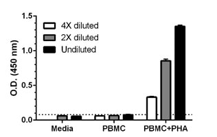 Comparison of secreted MIP3a in unstimulated and PHA-stimulated Human PBMC.
PBMC were grown in the absence or presence of phytohemagglutinin (PHA) for 2 days. MIP3a concentrations were measured in 4X and 2X diluted and undiluted (neat) cell culture supernatants of the stimulated PBMC, unstimulated PBMC and media. Raw data values (mean +/ SD, n=5) are graphed. The dotted line represents zero sample background.
-
 Interpolated concentrations of secreted MIP3a in unstimulated and PHA-stimulated Human PBMC.
The concentrations of MIP3a were interpolated from data values shown in Figure 3 using MIP3a standard curve and corrected for sample dilution. Using 4X and 2X diluted samples, the mean MIP3a concentration in stimulated PBMC supernatants was determined to be 1,151 pg/mL. The undiluted sample of stimulated PBMC supernatant was not considered into the calculation because of poor recovery of MIP3a in 100% media (data not shown). MIP3a concentrations in unstimulated PBMC supernatants measured less than the MDD, 39 pg/mL.


