Human Beta IG-H3 ELISA Kit (ab220651)
Key features and details
- One-wash 90 minute protocol
- Sensitivity: 6.3 pg/ml
- Range: 18.75 pg/ml - 1200 pg/ml
- Sample type: Cell culture extracts, Cell culture supernatant, Cit plasma, EDTA Plasma, Hep Plasma, Serum
- Detection method: Colorimetric
- Assay type: Sandwich (quantitative)
- Reacts with: Human
Overview
-
Product name
Human Beta IG-H3 ELISA Kit
See all TGFBI kits -
Detection method
Colorimetric -
Precision
Intra-assay Sample n Mean SD CV% Serum 8 2.4% Inter-assay Sample n Mean SD CV% Serum 3 3.2% -
Sample type
Cell culture supernatant, Serum, Cell culture extracts, Hep Plasma, EDTA Plasma, Cit plasma -
Assay type
Sandwich (quantitative) -
Sensitivity
6.3 pg/ml -
Range
18.75 pg/ml - 1200 pg/ml -
Recovery
Sample specific recovery Sample type Average % Range Cell culture supernatant 104 103% - 107% Serum 99 97% - 101% Cell culture extracts 94 92% - 98% Cell culture media 88 84% - 90% Hep Plasma 94 91% - 99% EDTA Plasma 101 98% - 105% Cit plasma 100 97% - 104% -
Assay time
1h 30m -
Assay duration
One step assay -
Species reactivity
Reacts with: Human
Does not react with: Cow -
Product overview
Human Beta IG-H3 ELISA Kit (ab220651) is a single-wash 90 min sandwich ELISA designed for the quantitative measurement of Beta IG-H3 protein in cell culture extracts, cell culture supernatant, serum, cit plasma, edta plasma, and hep plasma. It uses our proprietary SimpleStep ELISA® technology. Quantitate Human Beta IG-H3 with 6.3 pg/ml sensitivity.
SimpleStep ELISA® technology employs capture antibodies conjugated to an affinity tag that is recognized by the monoclonal antibody used to coat our SimpleStep ELISA® plates. This approach to sandwich ELISA allows the formation of the antibody-analyte sandwich complex in a single step, significantly reducing assay time. See the SimpleStep ELISA® protocol summary in the image section for further details. Our SimpleStep ELISA® technology provides several benefits:
- Single-wash protocol reduces assay time to 90 minutes or less
- High sensitivity, specificity and reproducibility from superior antibodies
- Fully validated in biological samples
- 96-wells plate breakable into 12 x 8 wells stripsA 384-well SimpleStep ELISA® microplate (ab203359) is available to use as an alternative to the 96-well microplate provided with SimpleStep ELISA® kits.
-
Notes
Human beta IG-H3 is also known as Kerato-epithelin and RGD-CAP (RGD-containing collagen associated protein), is a secreted adhesion molecule that binds to collagens and integrins via covalent and noncovalent interactions. It encodes a 683-amino acid protein containing a secretory signal sequence and four homologous internal domains. Mouse and bovine beta IG-H3 proteins are 90.6% and 92.7% homologous to human beta IG-H3, respectively.
Abcam has not and does not intend to apply for the REACH Authorisation of customers’ uses of products that contain European Authorisation list (Annex XIV) substances.
It is the responsibility of our customers to check the necessity of application of REACH Authorisation, and any other relevant authorisations, for their intended uses. -
Platform
Microplate (12 x 8 well strips)
Properties
-
Storage instructions
Store at +4°C. Please refer to protocols. -
Components 1 x 96 tests 10X Human beta IG-H3 Capture Antibody 1 x 600µl 10X Human beta IG-H3 Detector Antibody 1 x 600µl 10X Wash Buffer PT (ab206977) 1 x 20ml 50X Cell Extraction Enhancer Solution (ab193971) 1 x 1ml 5X Cell Extraction Buffer PTR (ab193970) 1 x 10ml Antibody Diluent CPI - HAMA Blocker (ab193969) 1 x 6ml Human beta IG-H3 Lyophilized Recombinant Protein 2 vials Plate Seals 1 unit Sample Diluent NS (ab193972) 1 x 50ml SimpleStep Pre-Coated 96-Well Microplate (ab206978) 1 unit Stop Solution 1 x 12ml TMB Development Solution 1 x 12ml -
Research areas
-
Function
Binds to type I, II, and IV collagens. This adhesion protein may play an important role in cell-collagen interactions. In cartilage, may be involved in endochondral bone formation. -
Tissue specificity
Highly expressed in the corneal epithelium. -
Involvement in disease
Defects in TGFBI are the cause of epithelial basement membrane corneal dystrophy (EBMD) [MIM:121820]; also known as Cogan corneal dystrophy or map-dot-fingerprint type corneal dystrophy. EBMD is a bilateral anterior corneal dystrophy characterized by grayish epithelial fingerprint lines, geographic map-like lines, and dots (or microcysts) on slit-lamp examination. Pathologic studies show abnormal, redundant basement membrane and intraepithelial lacunae filled with cellular debris. Although this disorder usually is not considered to be inherited, families with autosomal dominant inheritance have been identified.
Defects in TGFBI are the cause of corneal dystrophy Groenouw type 1 (CDGG1) [MIM:121900]; also known as corneal dystrophy granular type. Inheritance is autosomal dominant. Corneal dystrophies show progressive opacification of the cornea leading to severe visual handicap.
Defects in TGFBI are the cause of corneal dystrophy lattice type 1 (CDL1) [MIM:122200]. Inheritance is autosomal dominant.
Defects in TGFBI are a cause of corneal dystrophy Thiel-Behnke type (CDTB) [MIM:602082]; also known as corneal dystrophy of Bowman layer type 2 (CDB2).
Defects in TGFBI are the cause of Reis-Buecklers corneal dystrophy (CDRB) [MIM:608470]; also known as corneal dystrophy of Bowman layer type 1 (CDB1).
Defects in TGFBI are the cause of lattice corneal dystrophy type 3A (CDL3A) [MIM:608471]. CDL3A clinically resembles to lattice corneal dystrophy type 3, but differs in that its age of onset is 70 to 90 years. It has an autosomal dominant inheritance pattern.
Defects in TGFBI are the cause of Avellino corneal dystrophy (ACD) [MIM:607541]. ACD could be considered a variant of granular dystrophy with a significant amyloidogenic tendency. Inheritance is autosomal dominant. -
Sequence similarities
Contains 1 EMI domain.
Contains 4 FAS1 domains. -
Post-translational
modificationsGamma-carboxyglutamate residues are formed by vitamin K dependent carboxylation. These residues are essential for the binding of calcium. -
Cellular localization
Secreted > extracellular space > extracellular matrix. May be associated both with microfibrils and with the cell surface. - Information by UniProt
-
Alternative names
- RGD containing collagen associated protein
- AI181842
- AI747162
see all -
Database links
- Entrez Gene: 7045 Human
- Omim: 601692 Human
- SwissProt: Q15582 Human
- Unigene: 369397 Human
Images
-
SimpleStep ELISA technology allows the formation of the antibody-antigen complex in one single step, reducing assay time to 90 minutes. Add samples or standards and antibody mix to wells all at once, incubate, wash, and add your final substrate. See protocol for a detailed step-by-step guide.
-
Background-subtracted data values (mean +/- SD) are graphed.
-
Background-subtracted data values (mean +/- SD) are graphed.
-
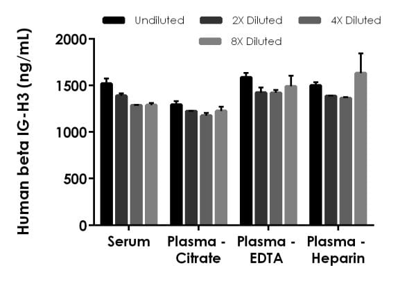
The concentrations of beta IG-H3 were measured in duplicates, interpolated from the beta IG-H3 standard curves and corrected for sample dilution. Undiluted samples are as follows: serum 1:4,000, plasma (citrate) 1:4,000, plasma (EDTA) 1:4,000, and plasma (heparin) 1:4,000. The interpolated dilution factor corrected values are plotted (mean +/- SD, n=2). The mean beta IG-H3 concentration was determined to be 1,371.41 ng/mL in neat serum, 1,229.89 ng/mL in neat plasma (citrate), 1,480.27 ng/mL in neat plasma (EDTA), and 1,470.88 ng/mL in neat plasma (heparin).
-
The concentrations of beta IG-H3 were measured in duplicates, interpolated from the beta IG-H3 standard curves and corrected for sample dilution. Undiluted samples are as follows: HUVEC supernatant 8%. The interpolated dilution factor corrected values are plotted (mean +/- SD, n=2). The mean beta IG-H3 concentration was determined to be 18,714.03 pg/mL in neat HUVEC supernatant. HUVEC supernatants were cultured in RPMI base media with 10% fetal bovine serum.
-
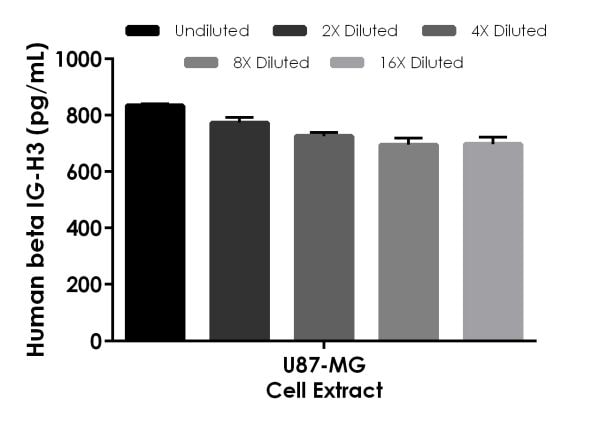
 Interpolated concentrations of native beta IG-H3 in human U87-MG cell extract based on a 10 µg/mL extract load.
The concentrations of beta IG-H3 were measured in duplicate and interpolated from the beta IG-H3 standard curve and corrected for sample dilution. The interpolated dilution factor corrected values are plotted (mean +/- SD, n=2). The mean beta IG-H3 concentration was determined to be 745.58 pg/mL in U87-MG cell extract.
-
Interpolated dilution factor corrected values are plotted (mean +/- SD, n=2). The mean beta IG-H3 concentration was determined to be 1,699.81 ng/mL with a range of 1,014.25 – 3,195.39 ng/mL.
-
To learn more about the advantages of recombinant antibodies see here.


