Goat Anti-Rabbit IgG H&L (FITC) (ab97050)
Key features and details
- Goat Anti-Rabbit IgG H&L (FITC)
- Conjugation: FITC. Ex: 493nm, Em: 528nm
- Host species: Goat
- Isotype: IgG
- Suitable for: IHC-P, ICC/IF, Flow Cyt
Overview
-
Product name
Goat Anti-Rabbit IgG H&L (FITC)
See all IgG secondary antibodies -
Host species
Goat -
Target species
Rabbit -
Specificity
By immunoelectrophoresis and ELISA this antibody reacts specifically with Rabbit IgG and with light chains common to other Rabbit immunoglobulins. No antibody was detected against non-immunoglobulin serum proteins. -
Tested applications
Suitable for: IHC-P, ICC/IF, Flow Cytmore details -
Immunogen
Full length protein. Rabbit IgG (containing the usual heavy and light chain)
-
Conjugation
FITC. Ex: 493nm, Em: 528nm
Properties
-
Form
Liquid -
Storage instructions
Shipped at 4°C. Store at +4°C. -
Storage buffer
pH: 6.8
Preservative: 0.09% Sodium azide
Constituents: 0.2% BSA, PBS -
 Concentration information loading...
 -
Purity
Immunogen affinity purified -
Purification notes
This antibody was isolated by affinity chromatography using antigen coupled to agarose beads and conjugated to FITC. -
Conjugation notes
F/P ratio is 4.85 -
Clonality
Polyclonal -
Isotype
IgG -
Research areas
Images
-
 Immunocytochemistry/ Immunofluorescence - Goat Anti-Rabbit IgG H&L (FITC) (ab97050) Ren Y et al. Potential of adipose-derived mesenchymal stem cells and skeletal muscle-derived satellite cells for somatic cell nuclear transfer mediated transgenesis in Arbas Cashmere goats. PLoS One 9:e93583 (2014).
Immunocytochemical/immunofluorescent analysis of 4% paraformaldehyde-fixed Triton X-100 permeabilized Goat skeletal muscle-derived satellite cells stained for NSE using ab53025.
After washing in PBS, the cells were incubated with Goat Anti-Rabbit IgG H&L (FITC) (ab97050) (Green).
-
 Flow Cytometry - Goat Anti-Rabbit IgG H&L (FITC) (ab97050) This image is courtesy of an anonymous Abreview.
ab84235 staining melanoma inhibitory activity in a human melanoma cell line by Flow Cytometry. Cells were harvested with EDTA and washed in PBS. Cells were pemeabilized with saponine. The sample was incubated with the primary antibody (1/100 in PBS) for 15 minutes at 20°C. A FITC-conjugated goat anti-rabbit IgG H&L (ab97050) (1/100) was used as the secondary antibody.
-
 Immunohistochemistry (Frozen sections) - Goat Anti-Rabbit IgG H&L (FITC) (ab97050) Courtesy of Dr. Shaohua Li, UMDNJ-Robert Wood Johnson Medical School
Sample: mouse embryonic stem cell-differentiated embryoid bodies (EBs)
Preparation:- Fix in 3% PFA in PBS for 30 min at RT
- Incubate in 7.5% sucrose-PBS for 3h at RT
- Incubate in 15% sucrose-PBS at 4 degree Celsius overnight
- Embed the EBs in tissue-Tek OCT compound
- Cut frozen sections to 4-20 µm thickness
Primary antibodies:
Mouse anti-Ki67, 1:100
Rabbit anti-laminin alpha 1 (basement marker)
Secondary antibodies:
Goat polyclonal Secondary Antibody to Mouse IgG - H&L (AMCA) (ab47052), 1:100
Goat polyclonal Secondary Antibody to Rabbit IgG - H&L (FITC) (ab97050), 1:100 -
-
-
-
-
-
The FACS staining was perform on THP-1 cell lines with Rabbit polyclonal to CCR2 (ab21667) at a 1/100 dillution for 30 min at 4C. The secondary antibody was ab97050 used at 1/100 for 20min at 4C. The buffer used was PBS/BSA (0.5%)/Azide (0.05%). No fixation or permeabilization was performed and gating was done on alive cells.
This image is courtesy of an anonymous Abreview
-
 Immunohistochemistry (Frozen sections) - Goat Anti-Rabbit IgG H&L (FITC) (ab97050) Courtesy of Dr. Shaohua Li, UMDNJ-Robert Wood Johnson Medical School
Sample: mouse embryonic stem cell-differentiated embryoid bodies (EBs)
Preparation:- Fix in 3%PFA in PBS for 30 min at RT
- Incubate in 7.5% sucrose-PBS for 3h at RT
- Incubate in 15% sucrose-PBS at 4 degree Celsius overnight
- Embed the EBs in tissue-Tek OCT compound
- Cut frozen sections to 4-20 µm thickness
Primary antibody: Rabbit anti-laminin alpha 1, 1:400
Secondary antibody: Goat polyclonal Secondary Antibody to Rabbit IgG - H&L (FITC) (ab97050)
F-actin was stained with CytoPainter F-actin staining kit (blue) (ab112124), 1:1000
Nuclei were counterstained stained with DRAQ7TM (ab109202), 1:1000 -
-
-
-
-
-
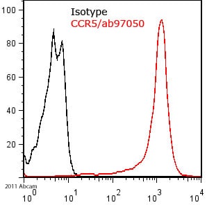
The FACS staining was performed on HEK cells expressing human CCR5 with PBS/BSA (0.5%)/Azide (0.05%) as FACS buffer. We used a primary polyclonal rabbit antibody against CCR5 at a 1/100 dillution, incubated for 1 hour at 4C. The secondary antibody was ab97050 used at a concentration of 1/100 for 30min at 4C. No fixation or permeabilization was performed and gaiting was done on alive cells.
This image is courtesy of an anonymous Abreview


