Embryonic Stem Cell Marker Panel (Mouse: Oct4, Nanog, SOX2, SSEA1) (ab107156)
Overview
-
Product name
Embryonic Stem Cell Marker Panel (Mouse: Oct4, Nanog, SOX2, SSEA1) -
Product overview
ab107156 is a Mouse Embryonic Stem Cell Marker Panel containing four antibodies: 50µg of Oct4 rabbit polyclonal, 50µl of Nanog rabbit polyclonal, 50µg of SOX2 rabbit polyclonal and 50µl of SSEA1 mouse monoclonal.
The Mouse Embryonic Stem Cell Marker Panel is designed for the validation and characterization of cultured or newly derived mouse embryonic stem (ES) cell lines. The panel contains four antibodies to well known markers of mouse ES cells (Oct4, Nanog, SOX2 and SSEA1). Embryonic stem cells are isolated from the inner cell mass of the early blastocyst. They have two properties that make them unique. Firstly, under defined conditions they can self-renew indefinitely and secondly they are pluripotent, which means they can differentiate into all three germ layers (ectoderm, mesoderm and endoderm).
The antibodies in this panel were selected for their exceptional performance in ICC/IF in mouse cells. In addition, these antibodies have also been tested in a number other applications and species. Please see the individual datasheets for additional information. Recommended working dilutions for ICC/IF in mouse can also be found in the figure legends on this datasheet.
-
Notes
Explore our range of antibody sample panels designed to provide you with a variety of trial-size antibodies in a convenient and cost-effective format.
Properties
-
Storage instructions
Please refer to protocols. -
Components 1 units ab16285 - Anti-SSEA1 antibody [MC-480] 1 x 50µl ab19857 - Anti-Oct4 antibody - ChIP Grade 1 x 50µg ab80892 - Anti-Nanog antibody 1 x 50µl ab97959 - Anti-SOX2 antibody 1 x 50µg -
Research areas
-
Alternative names
- Fucosyltransferase 4
- Fucosyltransferase IV
- Galactoside 3 L fucosyltransferase
see all
Images
-
 Immunocytochemistry/ Immunofluorescence - Mouse Embryonic Stem Cell Marker Panel (Oct4, Nanog, SOX2, SSEA1) (ab107156)ICC/IF image of Oct4 (ab19857, green) and SSEA1 (ab16285, red) co-stained mouse embryonic stem cells. The cells were fixed in 4% paraformaldehyde and then permeabilized in blocking buffer (0.1% BSA, 1% serum, 0.1% triton in PBS) for 30 minutes at 25°C. The cells were then incubated with Oct4 (1:250) and SSEA1 (1:10) overnight at 4°C. The secondary antibodies, alexa fluor 488 goat anti rabbit IgG (green) and alexa fluor 647 goat anti mouse IgM (red), were both use at a 1:500 dilution for 1 hour. DAPI was used to stain the cell nuclei (blue).
 -
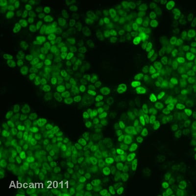
 Immunocytochemistry/ Immunofluorescence - Mouse Embryonic Stem Cell Marker Panel (Oct4, Nanog, SOX2, SSEA1) (ab107156)ab80892 staining Nanog (green) in mouse embryonic stem cells by immunocytochemistry/ immunofluorescence. Cells were paraformaldehyde fixed and permeabilized in blocking buffer (0.1% BSA, 1% serum, 0.1% triton in PBS) for 30 minutes at 25°C. The primary antibody was diluted 1:200 and incubated with the sample overnight at 4°C. An Alexa Fluor conjugated goat anti rabbit IgG antibody, diluted 1:500, was used as the secondary and incubated with samples for 1 hour at room temperature.
 -
 Immunocytochemistry/ Immunofluorescence - Mouse Embryonic Stem Cell Marker Panel (Oct4, Nanog, SOX2, SSEA1) (ab107156)ICC/IF image of Sox2 (ab97959, green) and SSEA1 (ab16285, red) co-stained mouse embryonic stem cells. The cells were fixed in paraformaldehyde and then permeabilized in blocking buffer (0.1% BSA, 1% serum, 0.1% triton in PBS) for 30 minutes at 25°C. The cells were then incubated with Sox2 (1:500) and SSEA1 (1:10) overnight at 4°C. The secondary antibodies, alexa fluor 488 goat anti rabbit IgG (green) and alexa fluor 647 goat anti mouse IgM (red), were both use at a 1:500 dilution for 1 hour at room temperature. DAPI was used to stain the cell nuclei (blue).
 -
 Immunocytochemistry/ Immunofluorescence - Mouse Embryonic Stem Cell Marker Panel (Oct4, Nanog, SOX2, SSEA1) (ab107156)ab16285 staining SSEA1 (red) in mouse embryonic stem cells by immunocytochemistry/ immunofluorescence. Cells were paraformaldehyde fixed and permeabilized in blocking buffer (0.1% BSA, 1% serum, 0.1% triton in PBS) for 30 minutes at 25°C. The primary antibody was diluted 1:10 and incubated with the sample overnight at 4°C. An Alexa Fluor 647 conjugated goat anti mouse IgM antibody, diluted 1:500, was used as the secondary and incubated with samples for 1 hour at room temperature.
 -


