CCCP, Mitochondrial oxidative phosphorylation uncoupler (ab141229)
Key features and details
- Potent mitochondrial oxidative phosphorylation uncoupler
- CAS Number: 555-60-2
- Purity: > 99%
- Soluble in DMSO to 100 mM and in ethanol to 100 mM
- Form / State: Solid
- Source: Synthetic
Overview
-
Product name
CCCP, Mitochondrial oxidative phosphorylation uncoupler -
Description
Potent mitochondrial oxidative phosphorylation uncoupler -
Biological description
Potent mitochondrial oxidative phosphorylation uncoupler. Renders mitochondrial inner membrane permeable to protons. Induces apoptosis in vitro.
-
Purity
> 99% -
CAS Number
555-60-2 -
Chemical structure

Properties
-
Chemical name
2-[2-(3-Chlorophenyl)hydrazinylyidene]propanedinitrile -
Molecular weight
204.62 -
Molecular formula
C9H5ClN4 -
PubChem identifier
2603 -
Storage instructions
Store at -20°C. Store under desiccating conditions. The product can be stored for up to 12 months. -
Solubility overview
Soluble in DMSO to 100 mM and in ethanol to 100 mM -
Handling
Wherever possible, you should prepare and use solutions on the same day. However, if you need to make up stock solutions in advance, we recommend that you store the solution as aliquots in tightly sealed vials at -20°C. Generally, these will be useable for up to one month. Before use, and prior to opening the vial we recommend that you allow your product to equilibrate to room temperature for at least 1 hour.
Toxic, refer to SDS for further information.
Need more advice on solubility, usage and handling? Please visit our frequently asked questions (FAQ) page for more details.
-
SMILES
Clc1cc(N\N=C(/C#N)C#N)ccc1 -
Source
Synthetic
-
Research areas
Images
-
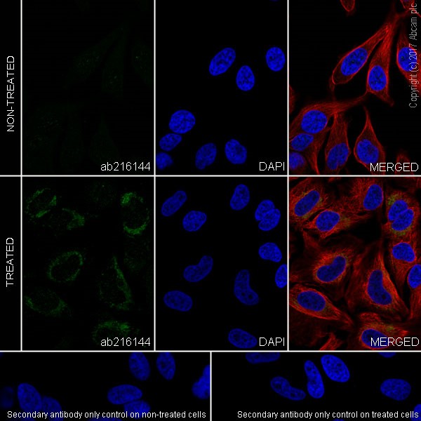
 Immunocytochemistry/ Immunofluorescence - CCCP, Mitochondrial oxidative phosphorylation uncoupler (ab141229)
Immunofluorescent analysis of 4 % paraformaldehyde-fixed, 0.1% Triton X-100 permeabilized HeLa (human epithelial cell line from cervix adenocarcinoma)(+/- treatment with 10μM carbonyl cyanide 3-chlorophenylhydrazone (CCCP, ab141229) for 24 hours) cells labeling PINK1 with ab216144 at 1/500 dilution, followed by Goat Anti-Rabbit IgG H&L (Alexa Fluor® 488) (ab150077) secondary antibody at 1/1000 dilution (green). Confocal image showing cytoplasmic staining on HeLa cells treated with 10μM carbonyl cyanide 3-chlorophenylhydrazone (CCCP, ab141229) for 24 hours. The nuclear counter stain is DAPI (blue). Tubulin is detected with Anti-alpha Tubulin antibody [DM1A] - Microtubule Marker (Alexa Fluor® 594) (ab195889) at 1/200 dilution (red).
The negative controls are as follows:
-ve control: PBS, followed by Goat Anti-Rabbit IgG H&L (Alexa Fluor® 488) (ab150077) secondary antibody at 1/1000 dilution. -
All lanes : Anti-PINK1 antibody [EPR20730] (ab216144) at 1/1000 dilution
Lane 1 : HeLa (human epithelial cell line from cervix adenocarcinoma) whole cell lysate
Lane 2 : HeLa cells (treated with 10uM carbonyl cyanide 3-chlorophenylhydrazone (CCCP, ab141229) for 24 hours) whole cell lysate
Lysates/proteins at 20 µg per lane.
Secondary
All lanes : Goat Anti-Rabbit IgG H&L (HRP) (ab97051) at 1/20000 dilution
Developed using the ECL technique.
Observed band size: 62 kDa why is the actual band size different from the predicted?
Exposure time: 5 secondsBlocking and dilution buffer: 5% NFDM/TBST
PINK1 can be induced by CCCP treatment (PMID: 24184327).
-
PINK1 was immunoprecipitated from 0.35 mg of HeLa (human epithelial cell line from cervix adenocarcinoma) (treated with 10uM carbonyl cyanide 3-chlorophenylhydrazone (CCCP. ab141229) for 24 hours) whole cell lysate with ab216144 at 1/30 dilution. Western blot was performed from the immunoprecipitate using ab216144 at 1/500 dilution. VeriBlot for IP Detection Reagent (HRP) (ab131366), was used for detection at 1/1000 dilution.
Lane 1: HeLa (CCCP-treated, ab141229) lysate 10 μg (Input).
Lane 2: ab216144 IP in HeLa (CCCP-treated, ab141229) lysate.
Lane 3: Rabbit monoclonal IgG (ab172730) instead of ab216144 in HeLa (CCCP-treated, ab141229) whole cell lysate.Blocking and dilution buffer: 5% NFDM/TBST.
-
MCF7 cells were incubated at 37°C for 2 hours with vehicle control (0 μM) and different concentrations of CCCP (ab 141229). Increased expression of AKT1 (phospho S473) (ab81283) in MCF7 cells correlates with an increase in CCCP concentration, as described in literature.
Whole cell lysates were prepared with RIPA buffer (containing protease inhibitors and sodium orthovanadate), 10 μg of each were loaded on the gel and the WB was run under reducing conditions. After transfer the membrane was blocked for an hour using 5% BSA before being incubated with ab81283 at 2 μg/ml and ab8227 at 1 μg/ml overnight at 4°C. Antibody binding was detected using an anti-rabbit antibody conjugated to HRP (ab97051) at 1/10000 and visualised using ECL development solution.


