APC Conjugation Kit - Lightning-Link® (ab201807)
Overview
-
Product name
APC Conjugation Kit - Lightning-Link® -
Product overview
APC Conjugation Kit / APC Labeling Kit (ab201807) uses a simple and quick process for APC labeling / conjugation of antibodies. It can also be used to conjugate other proteins or peptides. Learn about our antibody labeling kits and their advantages.
To conjugate an antibody to APC using this kit:
- add modifier to antibody and incubate for 3 hours
- add quencher and incubate for 30 mins
The APC conjugated antibody can be used immediately in WB, ELISA, IHC etc. No further purification is required and 100% of the antibody is recovered for use.Learn about buffer compatibility below; for incompatible buffers and low antibody concentrations, use our rapid antibody purification and concentration kits. Use the FAQ to learn more about the technology, or about conjugating other proteins and peptides to APC.
Custom size conjugation kits up to 100 mg are available on demand. Please contact us to discuss your requirements.
-
Notes
This product is manufactured by Expedeon, an Abcam company, and was previously called Lightning-Link® APC Labeling Kit. 705-0015 is the same as the 1 mg size. 705-0010 is the same as the 3 x 100 ug size. 705-0030 is the same as the 3 x 30 ug size. 705-0005 is the same as the 100 µg size.
Amount and volume of antibody for conjugation to APC
Kit size Recommended
maximum amount of antibodyMaximum antibody
volume3 x 10 µg 3 x 10 µg 3 x 10 µL 100 µg 1x 100 µg 1 x 100 µL 3 x 100 µg 3 x 100 µg 3 x 100 µL 1 mg 1 x 1 mg 1 x 1 mL 1 Ideal antibody concentration is 1mg/ml. 0.5 - 1 mg/ml can be used if the maximum antibody volume is not exceeded. Antibodies > 1 mg/ml or
Buffer Requirements for Conjugation
Buffer should be pH 6.5-8.5.
Compatible buffer constituents
If a concentration is shown, then the constituent should be no more than the concentration shown. If several constituents are close to the limit of acceptable concentration, then this can inhibit conjugation.50mM / 0.6% Tris1 0.1% BSA2 50% glycerol 0.1% sodium azide PBS Potassium phosphate Sodium chloride HEPES Sucrose Sodium citrate EDTA Trehalose 1 Tris buffered saline is almost always ≤ 50 mM / 0.6%
2 BSA can also interfere with the use of the conjugated antibody in tissue staining.Incompatible buffer constituents
Thiomerosal Proclin Glycine Arginine Glutathione DTT If a constituent of the buffer containing your antibody or protein is not listed above, please check the FAQ or contact us.
Only purified antibodies are suitable for use, ie. where other proteins, peptides, or amino acids are not present: antibodies in ascites fluid, serum or hybridoma culture media are incompatible.
Properties
-
Storage instructions
Store at -20°C. Please refer to protocols. -
Components 100 µg 1 mg 3 x 10 µg 3 x 100 µg ab274126 - APC Conjugation Mix 1 x 100µg 1 x 1mg 3 x 10µg 3 x 100µg ab274106 - Modifier reagent 1 x 200µl 1 x 200µl 1 x 200µl 1 x 200µl ab274296 - Quencher reagent 1 x 200µl 1 x 200µl 1 x 200µl 1 x 200µl -
Research areas
-
Alternative names
- Allophycocyanin
Images
-
-
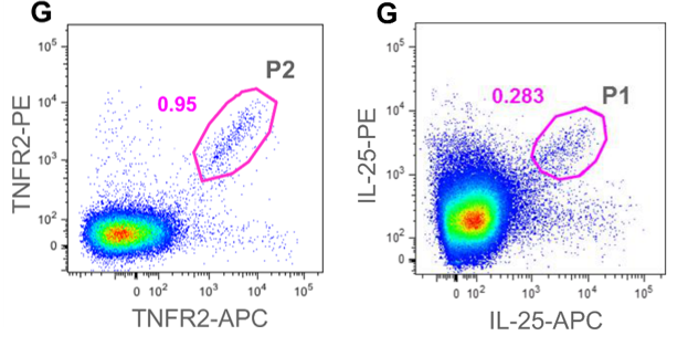
 APC Conjugation Kit - Lightning-Link® labeling IL-25 and TNFR2 extracellular domain for Flow cytometry Image from Starkie DO et al., PLoS One, 11(3):e0152282. Fig 3 and 2.; doi: 10.1371/journal.pone.0152282. Reproduced under the Creative Commons license https://creativecommons.org/licenses/by/4.0/
Starkie DO et al. used ab201807 to identify antigen-specific mouse memory B cells from TNFR2 and IL-25 immunised mice.
-
 APC Conjugation Kit - Lightning-Link® labeling anti-human 4ß7 antibody for Flow cytometry Image from McLinden RJ et al., PLoS One, 8(11):e77756. Fig 2 .; doi: 10.1371/journal.pone.0077756. Reproduced under the Creative Commons license https://creativecommons.org/licenses/by/4.0/
McLinden RJ et al. used ab201807 as part of examining HIV-1 neutralizing antibodies.
They used the kit to conjugate APC to anti-human 4β7 antibody for use in flow cytometry.
A. Flow cytometric analysis of CD4, CCR5 and α4β7 expression in the A3R5.7 cell line. 0.5 x 10⁶ cells were singly stained for 30 minutes with fluorochrome-conjugated antibodies as shown followed by fixation in 2% paraformaldehyde. Data are representative of at least two independent experiments. Isotype controls are shown in grey. Nearly all cells were positive for CD4 and CCR5 while approximately half were positive for α4β7.
B. Comparison of cell surface CD4, CCR5 and α4β7 receptor densities in various cell targets. 0.5 x 10⁶ cells were stained with fluorochrome-conjugated antibodies and compared to defined populations of similarly stained Quantum Simply Cellular beads. PBMC were stimulated with CD3.8 bi-specific antibody in the presence of 50U/mL rhIL-2. Assuming monovalent antibody-to-surface receptor binding, the Antibody Binding Capacity (ABC) calculated is equivalent to receptors/cell. Data represents the mean of two separate experiments. TZM-bl cells express high levels of CD4 and CCR5 but are negative for α4β7 while A3R5.7 cells possess CCR5 and α4β7 densities more similar to PBMC. CD4 expression on TZM-bl was beyond assay range.
-
 APC Conjugation Kit - Lightning-Link® labeling OVA and OVA-CRT for in-gel Fluorescence and Flow cytometry Image from Del Cid N et al., PLoS One, 7(7):e41727. Fig 2.; doi: 10.1371/journal.pone.0041727. Reproduced under the Creative Commons license https://creativecommons.org/licenses/by/4.0/
Del Cid N et al. used ab201807 as part of examining antigen cross-presentation.
They used the kit to conjugate APC to ovalbumin (OVA) and ovalbumin-calreticulin fusion protein (OVA-CRT) for use in in-gel fluorescence and flow cytometry.
OVA-CRT and OVA were labeled with allophycocyanin. (C) Labeling intensity was determined by fluorescence imaging of the proteins after separation by SDS-PAGE (inset). Fluorescence intensity was quantified for the indicated proteins. (D) Binding of fluorescent proteins to BMDC was assessed by flow cytometry. BMDC were incubated with labeled proteins on ice before being analyzed by flow cytometry. BMDC not incubated with proteins are depicted as a grey filled. -
 APC Conjugation Kit - Lightning-Link® labeling anti-mouse glucagon antibody for Flow cytometry Image from Kalis M et al., PLoS One, 6(12):e29166. Fig 5.; doi: 10.1371/journal.pone.0029166. Reproduced under the Creative Commons license https://creativecommons.org/licenses/by/4.0/


