Anti-WWP1 antibody (ab43791)
Key features and details
- Rabbit polyclonal to WWP1
- Suitable for: ICC, WB
- Reacts with: Human
- Isotype: IgG
Overview
-
Product name
Anti-WWP1 antibody
See all WWP1 primary antibodies -
Description
Rabbit polyclonal to WWP1 -
Host species
Rabbit -
Tested applications
Suitable for: ICC, WBmore details -
Species reactivity
Reacts with: Human
Predicted to work with: Mouse, Rat, Cow
-
Immunogen
Synthetic peptide corresponding to Human WWP1 aa 200-300 conjugated to keyhole limpet haemocyanin.
(Peptide available asab43790) -
Positive control
- WB: HeLa and MCF7 whole cell lysate. ICC: A431 cells.
-
General notes
The Life Science industry has been in the grips of a reproducibility crisis for a number of years. Abcam is leading the way in addressing this with our range of recombinant monoclonal antibodies and knockout edited cell lines for gold-standard validation. Please check that this product meets your needs before purchasing.
If you have any questions, special requirements or concerns, please send us an inquiry and/or contact our Support team ahead of purchase. Recommended alternatives for this product can be found below, along with publications, customer reviews and Q&As
Properties
-
Form
Liquid -
Storage instructions
Shipped at 4°C. Store at +4°C short term (1-2 weeks). Upon delivery aliquot. Store at -20°C or -80°C. Avoid freeze / thaw cycle. -
Storage buffer
pH: 7.40
Preservative: 0.02% Sodium azide
Constituent: PBS
Batches of this product that have a concentration Concentration information loading...
Purity
Immunogen affinity purifiedClonality
PolyclonalIsotype
IgGResearch areas
Associated products
-
Compatible Secondaries
-
Isotype control
-
Recombinant Protein
Applications
The Abpromise guarantee
Our Abpromise guarantee covers the use of ab43791 in the following tested applications.
The application notes include recommended starting dilutions; optimal dilutions/concentrations should be determined by the end user.
Application Abreviews Notes ICC Use a concentration of 1 µg/ml.WB Use a concentration of 1 µg/ml. Detects a band of approximately 105 kDa (predicted molecular weight: 105 kDa).Notes ICC
Use a concentration of 1 µg/ml.WB
Use a concentration of 1 µg/ml. Detects a band of approximately 105 kDa (predicted molecular weight: 105 kDa).Target
-
Relevance
WWP1 is an E3 ubiquitin ligase and belongs to a family of NEDD4-like proteins. WWP1 contains 4 tandem WW domains and a HECT (homologous to the E6-associated protein carboxyl terminus) domain. WW domain-containing proteins are found in all eukaryotes and play an important role in the regulation of a wide variety of cellular functions such as protein degradation, transcription, and RNA splicing. The HECT domain of WWP1 has been implicated in regulating the localization and stability of p53 – inhibition of WWP1 results in a decrease in p53 expression, whilst WWP1 mediated stabilization of p53 appears to be associated with an accumulation of cytoplasmic p53. WWP1 also negatively regulates the TGF beta tumor suppressor pathway by inactivating its molecular components (SMAD2, SMAD4 and TGFbetaR1). WWP1 has been implicated in both breast and prostate cancers. -
Cellular localization
Cell Membrane, Cytoplasmic and Nuclear -
Database links
- Entrez Gene: 513789 Cow
- Entrez Gene: 11059 Human
- Entrez Gene: 107568 Mouse
- Entrez Gene: 297930 Rat
- Omim: 602307 Human
- SwissProt: Q9H0M0 Human
- SwissProt: Q8BZZ3 Mouse
-
Alternative names
- AIP 5 antibody
- AIP5 antibody
- Atropin-1-interacting protein 5 antibody
see all
Images
-
ab43791 staining WWP1 in A431 cells. The cells were fixed with 4% paraformaldehyde (10 min), permeabilized with 0.1% PBS-Tween for 5 minutes and then blocked with 1% BSA/10% normal goat serum/0.3M glycine in 0.1%PBS-Tween for 1h. The cells were then incubated overnight at 4°C with ab43791 at 1µg/ml and ab7291, Mouse monoclonal [DM1A] to alpha Tubulin - Loading Control. Cells were then incubated with ab150081, Goat polyclonal Secondary Antibody to Rabbit IgG - H&L (Alexa Fluor® 488), pre-adsorbed at 1/1000 dilution (shown in green) and ab150120, Goat polyclonal Secondary Antibody to Mouse IgG - H&L (Alexa Fluor® 594), pre-adsorbed at 1/1000 dilution (shown in pseudocolour red). Nuclear DNA was labelled with DAPI (shown in blue).
Image was acquired with a confocal microscope (Leica-Microsystems TCS SP8) and a single confocal section is shown.
-
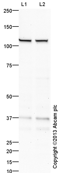
All lanes : Anti-WWP1 antibody (ab43791) at 1 µg/ml
Lane 1 : MCF7 (Human breast adenocarcinoma cell line) Whole Cell Lysate
Lane 2 : HeLa (Human epithelial carcinoma cell line) Whole Cell Lysate
Lysates/proteins at 10 µg per lane.
Secondary
All lanes : Goat polyclonal to Rabbit IgG - H&L - Pre-Adsorbed (HRP) at 1/50000 dilution
Developed using the ECL technique.
Performed under reducing conditions.
Predicted band size: 105 kDa
Observed band size: 105 kDa
Additional bands at: 36 kDa. We are unsure as to the identity of these extra bands.
Exposure time: 8 minutesThis blot was produced using a 4-12% Bis-tris gel under the MOPS buffer system. The gel was run at 200V for 50 minutes before being transferred onto a Nitrocellulose membrane at 30V for 70 minutes. The membrane was then blocked for an hour using 2% Bovine Serum Albumin before being incubated with ab43791 overnight at 4°C. Antibody binding was detected using an anti-rabbit antibody conjugated to HRP, and visualised using ECL development solution ab133406.
Protocols
Datasheets and documents
-
SDS download
-
Datasheet download
References (9)
ab43791 has been referenced in 9 publications.
- Jing Y et al. Mutant NPM1-regulated lncRNA HOTAIRM1 promotes leukemia cell autophagy and proliferation by targeting EGR1 and ULK3. J Exp Clin Cancer Res 40:312 (2021). PubMed: 34615546
- Zhong G et al. WWP1 Deficiency Alleviates Cardiac Remodeling Induced by Simulated Microgravity. Front Cell Dev Biol 9:739944 (2021). PubMed: 34733849
- Kishikawa T et al. WWP1 inactivation enhances efficacy of PI3K inhibitors while suppressing their toxicities in breast cancer models. J Clin Invest 131:N/A (2021). PubMed: 34907909
- Li Y et al. MicroRNA-15b shuttled by bone marrow mesenchymal stem cell-derived extracellular vesicles binds to WWP1 and promotes osteogenic differentiation. Arthritis Res Ther 22:269 (2020). PubMed: 33198785
- Lee YR et al. Reactivation of PTEN tumor suppressor for cancer treatment through inhibition of a MYC-WWP1 inhibitory pathway. Science 364:N/A (2019). PubMed: 31097636
- Baillet N et al. E3 Ligase ITCH Interacts with the Z Matrix Protein of Lassa and Mopeia Viruses and Is Required for the Release of Infectious Particles. Viruses 12:N/A (2019). PubMed: 31906112
- Matsudaira T et al. Endosomal phosphatidylserine is critical for the YAP signalling pathway in proliferating cells. Nat Commun 8:1246 (2017). PubMed: 29093443
- Goto Y et al. Regulation of E3 ubiquitin ligase-1 (WWP1) by microRNA-452 inhibits cancer cell migration and invasion in prostate cancer. Br J Cancer 114:1135-44 (2016). PubMed: 27070713
- Anindya R et al. Damage-induced ubiquitylation of human RNA polymerase II by the ubiquitin ligase Nedd4, but not Cockayne syndrome proteins or BRCA1. Mol Cell 28:386-97 (2007). PubMed: 17996703
Images
-
All lanes : Anti-WWP1 antibody (ab43791) at 1 µg/ml
Lane 1 : MCF7 (Human breast adenocarcinoma cell line) Whole Cell Lysate
Lane 2 : HeLa (Human epithelial carcinoma cell line) Whole Cell Lysate
Lysates/proteins at 10 µg per lane.
Secondary
All lanes : Goat polyclonal to Rabbit IgG - H&L - Pre-Adsorbed (HRP) at 1/50000 dilution
Developed using the ECL technique.
Performed under reducing conditions.
Predicted band size: 105 kDa
Observed band size: 105 kDa
Additional bands at: 36 kDa. We are unsure as to the identity of these extra bands.
Exposure time: 8 minutesThis blot was produced using a 4-12% Bis-tris gel under the MOPS buffer system. The gel was run at 200V for 50 minutes before being transferred onto a Nitrocellulose membrane at 30V for 70 minutes. The membrane was then blocked for an hour using 2% Bovine Serum Albumin before being incubated with ab43791 overnight at 4°C. Antibody binding was detected using an anti-rabbit antibody conjugated to HRP, and visualised using ECL development solution ab133406.
-
ICC/IF image of ab43791 stained HeLa cells. The cells were 4% formaldehyde fixed (10 min) and then incubated in 1%BSA / 10% normal goat serum / 0.3M glycine in 0.1% PBS-Tween for 1h to permeabilise the cells and block non-specific protein-protein interactions. The cells were then incubated with the antibody (ab43791, 5µg/ml) overnight at +4°C. The secondary antibody (green) was Alexa Fluor® 488 goat anti-rabbit IgG (H+L) used at a 1/1000 dilution for 1h. Alexa Fluor® 594 WGA was used to label plasma membranes (red) at a 1/200 dilution for 1h. DAPI was used to stain the cell nuclei (blue) at a concentration of 1.43µM. This antibody also gave a positive result in 4% formaldehyde fixed (10 min) Hek293 and HepG2 cells at 5µg/ml, and in 100% methanol fixed (5 min) HeLa, Hek293, HepG2 and MCF7 cells at 5µg/ml.


