Anti-USP9x antibody (ab19879)
Key features and details
- Rabbit polyclonal to USP9x
- Suitable for: IP, ICC, WB, IHC (PFA fixed)
- Knockout validated
- Reacts with: Mouse, Rat, Human
- Isotype: IgG
Overview
-
Product name
Anti-USP9x antibody
See all USP9x primary antibodies -
Description
Rabbit polyclonal to USP9x -
Host species
Rabbit -
Specificity
ab19879 detects 289kDa full length USP9X Human protein (Q93008) in WB on Caco2 Lysate. All detected bands are quenched by the immunizing peptide ab20617 -
Tested Applications & Species
See all applications and species dataApplication Species ICC HumanIHC (PFA fixed) RatIP HumanWB MouseHuman -
Immunogen
Synthetic peptide conjugated to KLH derived from within residues 1 - 100 of Human USP9x.
Read Abcam's proprietary immunogen policy -
Positive control
- ICC: Hek293 cells
Images
-
ab19879 staining USP9x in Hek293 cells. The cells were fixed with 100% methanol (5 min), permeabilized with 0.1% PBS-Triton X-100 for 5 minutes and then blocked with 1% BSA/10% normal goat serum/0.3M glycine in 0.1%PBS-Tween for 1h. The cells were then incubated overnight at 4°C with ab19879 at 1µg/ml and ab7291, Mouse monoclonal [DM1A] to alpha Tubulin - Loading Control. Cells were then incubated with ab150081, Goat polyclonal Secondary Antibody to Rabbit IgG - H&L (Alexa Fluor® 488), pre-adsorbed at 1/1000 dilution (shown in green) and ab150080, Goat polyclonal Secondary Antibody to Rabbit IgG - H&L (Alexa Fluor® 594) at 1/1000 dilution (shown in pseudocolour red). Nuclear DNA was labelled with DAPI (shown in blue). Also suitable in cells fixed with 4% paraformaldehyde (10 min).
Image was acquired with a high-content analyser (Operetta CLS, Perkin Elmer) and a maximum intensity projection of confocal sections is shown.
-
All lanes : Anti-USP9x antibody (ab19879) at 1 µg/ml
Lane 1 : Wild-type HeLa cell lysate
Lane 2 : USP9X knockout HeLa cell lysate
Lysates/proteins at 20 µg per lane.
Performed under reducing conditions.
Predicted band size: 54, 100,105 , 290 kDa
Observed band size: 290 kDa why is the actual band size different from the predicted?Lanes 1- 2: Merged signal (red and green). Green - ab19879 observed at 290 kDa. Red - Anti-alpha Tubulin antibody [DM1A] - Loading Control (ab7291) observed at 50 kDa.
ab19879 was shown to react with USP9x in wild-type HeLa cells in western blot. Loss of signal was observed when knockout cell line ab265665 (knockout cell lysate ab257790) was used. Wild-type HeLa and USP9X knockout HeLa cell lysates were subjected to SDS-PAGE. Membrane was blocked for 1 hour at room temperature in 0.1% TBST with 3% non-fat dried milk. ab19879 and Anti-alpha Tubulin antibody [DM1A] - Loading Control (ab7291) were incubated overnight at 4°C at a 1 µg/ml and a 1 in 20000 dilution respectively. Blots were developed with Goat anti-Rabbit IgG H&L (IRDye®800CW) preadsorbed (ab216773) and Goat anti-Mouse IgG H&L (IRDye®680RD) preadsorbed (ab216776) secondary antibodies at 1 in 20000 dilution for 1 hour at room temperature before imaging.
-
 Immunohistochemistry (PFA fixed) - Anti-USP9x antibody (ab19879) This image is courtesy of Sophie Pezet, King's College London, United Kingdom
Immuofluorescent staining for USP9X in the rat hippocampus (dentate gyrus) using ab19879 (1/300 = 0.07µg/ml). Image is taken with X10 objective. ab19879 was incubated overnight at RT. Secondary antibody used was anti-rabbit Alexa fluor 488 (1/1000 for 2h at RT). Rats were intracardially perfused with paraformaldehyde 4%, brain tissue was post-fixed overnight in the same fixative, cryoprotected in 20% sucrose and frozen in OCT. 30µm coronal sections were cut on a cryostat for free floating IHC.
-
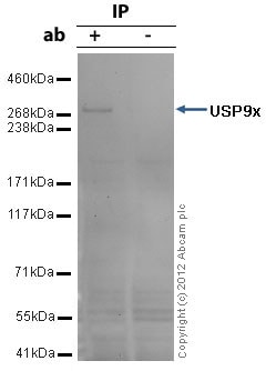
USP9x was immunoprecipitated using 0.5mg Caco2 whole cell extract, 5µg of Rabbit polyclonal to USP9x and 50µl of protein G magnetic beads (+). No antibody was added to the control (-).
The antibody was incubated under agitation with Protein G beads for 10min, Caco2 whole cell extract lysate diluted in RIPA buffer was added to each sample and incubated for a further 10min under agitation.
Proteins were eluted by addition of 40µl SDS loading buffer and incubated for 10min at 70oC; 10µl of each sample was separated on a SDS PAGE gel, transferred to a nitrocellulose membrane, blocked with 5% BSA and probed with ab19879.
Secondary: Mouse monoclonal [SB62a] Secondary Antibody to Rabbit IgG light chain (HRP) (ab99697).
Band: 290kDa: USP9x. -
Lane 1: Wild-type HAP1 cell lysate (20 µg)
Lane 2: USP9x knockout HAP1 cell lysate (20 µg)
Lane 3: T84 cell lysate (20 µg)
Lane 4: NIH3T3 cell lysate (20 µg)
Lanes 1 to 4: Merged signal (red and green). Green - ab19879 observed at 290 kDa. Red - loading control, ab181602 observed at 124 kDa.
ab19879 was shown to specifically react with USP9x when USP9x knockout samples were used. Wild-type and USP9x knockout samples were subjected to SDS-PAGE. ab9879 and ab181602 (loading control to GAPDH) were both diluted at 1 µg/ml and 1/10000 respectively and incubated overnight at 4°C. Blots were developed withGoat anti-Mouse IgG H&L (IRDye® 800CW) preadsorbed (ab216772) and Goat Anti-Rabbit IgG H&L (IRDye® 680RD) preadsorbed (ab216777) secondary antibodies at 1/10000 dilution for 1 h at room temperature before imaging. -
All lanes : Anti-USP9x antibody (ab19879) at 1 µg/ml
Lane 1 :Caco-2 whole cell lysate (ab3950)
Lane 2 :Caco-2 whole cell lysate (ab3950) with Human USP9x peptide (ab20617) at 1 µg/ml
Lysates/proteins at 20 µg per lane.
Secondary
All lanes : Alexa Fluor Goat polyclonal to Rabbit IgG (700) at 1/5000 dilution
Developed using the ECL technique.
Performed under reducing conditions.
Predicted band size: 54, 100,105 , 290 kDa
Observed band size: 100,105,290 kDa why is the actual band size different from the predicted?
Additional bands at: ~50-65 kDa. We are unsure as to the identity of these extra bands.ab19879 detects full length USP9x protein as well as a number of USP9x fragments in WB on Caco2 Lysate:
289kDa Human protein: Q93008 USP9X (Full length protein)
105kDa Human protein: Q6P468 - USP9X protein (Fragment) Human protein
99.7kDa Human protein: Q59EZ5 - USP9X protein variant (Fragment).
53.9kDa Q86X58 - USP9X protein (Fragment).


