Anti-USP10 antibody (ab70895)
")
Key features and details
- Rabbit polyclonal to USP10
- Suitable for: IHC-P, WB, IP, ICC/IF
- Reacts with: Mouse, Human
- Isotype: IgG
Overview
-
Product name
Anti-USP10 antibody
See all USP10 primary antibodies -
Description
Rabbit polyclonal to USP10 -
Host species
Rabbit -
Tested applications
Suitable for: IHC-P, WB, IP, ICC/IFmore details -
Species reactivity
Reacts with: Mouse, Human
Predicted to work with: Rhesus monkey, Orangutan
-
Immunogen
A region between residue 50 and 100 of human USP10 (NP_005144.1).
-
Positive control
- Whole cell lysate from HeLa and 293T cells.
-
General notes
Reproducibility is key to advancing scientific discovery and accelerating scientists’ next breakthrough.
Abcam is leading the way with our range of recombinant antibodies, knockout-validated antibodies and knockout cell lines, all of which support improved reproducibility.
We are also planning to innovate the way in which we present recommended applications and species on our product datasheets, so that only applications & species that have been tested in our own labs, our suppliers or by selected trusted collaborators are covered by our Abpromise™ guarantee.
In preparation for this, we have started to update the applications & species that this product is Abpromise guaranteed for.
We are also updating the applications & species that this product has been “predicted to work with,” however this information is not covered by our Abpromise guarantee.
Applications & species from publications and Abreviews that have not been tested in our own labs or in those of our suppliers are not covered by the Abpromise guarantee.
Please check that this product meets your needs before purchasing. If you have any questions, special requirements or concerns, please send us an inquiry and/or contact our Support team ahead of purchase. Recommended alternatives for this product can be found below, as well as customer reviews and Q&As.
Properties
-
Form
Liquid -
Storage instructions
Shipped at 4°C. Upon delivery aliquot and store at -20°C. Avoid freeze / thaw cycles. -
Storage buffer
pH: 6.8
Preservative: 0.09% Sodium azide
Constituents: 0.1% BSA, Tris buffered saline -
 Concentration information loading...
 -
Purity
Immunogen affinity purified -
Clonality
Polyclonal -
Isotype
IgG -
Research areas
Images
-
Western blot - Anti-USP10 antibody (ab70895)All lanes : Anti-USP10 antibody (ab70895) at 0.04 µg/ml
Lane 1 : Whole cell lysate from HeLa cells at 50 µg
Lane 2 : Whole cell lysate from HeLa cells at 15 µg
Lane 3 : Whole cell lysate from HeLa cells at 5 µg
Lane 4 : Whole cell lysate from 293T cells at 50 µg
Predicted band size: 87 kDa
Observed band size: 105 kDa why is the actual band size different from the predicted?
-
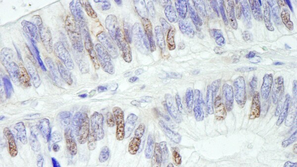 Immunohistochemistry (Formalin/PFA-fixed paraffin-embedded sections) - Anti-USP10 antibody (ab70895)Immunohistochemistry (Formalin/PFA-fixed paraffin-embedded sections) analysis of human colon carcinoma tissue labelling USP10 with ab70895 at 1/1000 (0.2µg/ml). Detection: DAB.
 -
 Immunoprecipitation - Anti-USP10 antibody (ab70895)Detection of Human USP10 by Immunoprecipitation in Whole cell lysate from HeLa cells (1 mg for IP, 20% of IP loaded), using ab70895 at 3 µg/mg lysate for IP, and at 1 µg/ml for subsequent Western blot detection.
 -
 Western blot - Anti-USP10 antibody (ab70895)All lanes : Anti-USP10 antibody (ab70895) at 0.1 µg/ml
Lane 1 : TCMK-1 whole cell lysate
Lane 2 : 4T1 whole cell lysate
Lane 3 : CT26.WT whole cell lysate
Lysates/proteins at 50 µg per lane.
Developed using the ECL technique.
Predicted band size: 87 kDa
Exposure time: 3 minutes
-
 Immunocytochemistry/ Immunofluorescence - Anti-USP10 antibody (ab70895)ICC/IF image of ab70895 stained HeLa cells. The cells were 4% formaldehyde fixed (10 min) and then incubated in 1%BSA / 10% normal goat serum / 0.3M glycine in 0.1% PBS-Tween for 1h to permeabilise the cells and block non-specific protein-protein interactions. The cells were then incubated with the antibody (ab70895, 1µg/ml) overnight at +4°C. The secondary antibody (green) was Alexa Fluor® 488 goat anti-rabbit IgG (H+L) used at a 1/1000 dilution for 1h. Alexa Fluor® 594 WGA was used to label plasma membranes (red) at a 1/200 dilution for 1h. DAPI was used to stain the cell nuclei (blue) at a concentration of 1.43µM.
-
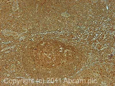 Immunohistochemistry (Formalin/PFA-fixed paraffin-embedded sections) - Anti-USP10 antibody (ab70895)IHC image of ab70895 staining in human normal tonsil formalin fixed paraffin embedded tissue section, performed on a Leica BondTM system using the standard protocol F. The section was pre-treated using heat mediated antigen retrieval with sodium citrate buffer (pH6, epitope retrieval solution 1) for 20 mins. The section was then incubated with ab70895, 5µg/ml, for 15 mins at room temperature and detected using an HRP conjugated compact polymer system. DAB was used as the chromogen. The section was then counterstained with haematoxylin and mounted with DPX.
For other IHC staining systems (automated and non-automated) customers should optimize variable parameters such as antigen retrieval conditions, primary antibody concentration and antibody incubation times.


