Anti-UAP56 antibody (ab47955)
")
Key features and details
- Rabbit polyclonal to UAP56
- Suitable for: IHC-P, WB, ICC/IF
- Reacts with: Human
- Isotype: IgG
Overview
-
Product name
Anti-UAP56 antibody
See all UAP56 primary antibodies -
Description
Rabbit polyclonal to UAP56 -
Host species
Rabbit -
Tested applications
Suitable for: IHC-P, WB, ICC/IFmore details -
Species reactivity
Reacts with: Human
Predicted to work with: Mouse, Rat, Chicken, Cow, Pig, Chimpanzee
-
Immunogen
Synthetic peptide conjugated to KLH derived from within residues 300 - 400 of Human UAP56.
Read Abcam's proprietary immunogen policy (Peptide available as ab48225.) -
Positive control
- ab47955 gave a positive signal in the following Whole Cell Lysates: Hela, Jurkat, HepG2 and A431. It also gave a positive signal in human tonsil tissue sections.
Properties
-
Form
Liquid -
Storage instructions
Shipped at 4°C. Store at +4°C short term (1-2 weeks). Upon delivery aliquot. Store at -20°C or -80°C. Avoid freeze / thaw cycle. -
Storage buffer
pH: 7.40
Preservative: 0.02% Sodium azide
Constituent: PBS
Batches of this product that have a concentration Concentration information loading...
Purity
Immunogen affinity purifiedClonality
PolyclonalIsotype
IgGResearch areas
Associated products
-
Compatible Secondaries
-
Isotype control
-
Recombinant Protein
Applications
Our Abpromise guarantee covers the use of ab47955 in the following tested applications.
The application notes include recommended starting dilutions; optimal dilutions/concentrations should be determined by the end user.
Application Abreviews Notes IHC-P Use a concentration of 1 µg/ml. Perform heat mediated antigen retrieval before commencing with IHC staining protocol. WB Use a concentration of 1 µg/ml. Detects a band of approximately 49 kDa (predicted molecular weight: 49 kDa). ICC/IF Use a concentration of 1 µg/ml. Target
-
Function
Component of the THO subcomplex of the TREX complex. The TREX complex specifically associates with spliced mRNA and not with unspliced pre-mRNA. It is recruited to spliced mRNAs by a transcription-independent mechanism. Binds to mRNA upstream of the exon-junction complex (EJC) and is recruited in a splicing- and cap-dependent manner to a region near the 5' end of the mRNA where it functions in mRNA export. The recruitment occurs via an interaction between THOC4 and the cap-binding protein NCBP1. DDX39B functions as a bridge between THOC4 and the THO complex. The TREX complex is essential for the export of Kaposi's sarcoma-associated herpesvirus (KSHV) intronless mRNAs and infectious virus production. The recruitment of the TREX complex to the intronless viral mRNA occurs via an interaction between KSHV ORF57 protein and THOC4.
Splice factor that is required for the first ATP-dependent step in spliceosome assembly and for the interaction of U2 snRNP with the branchpoint. Has both RNA-stimulated ATP binding/hydrolysis activity and ATP-dependent RNA unwinding activity. Even with the stimulation of RNA, the ATPase activity is weak. Can only hydrolyze ATP but not other NTPs. The RNA stimulation of ATPase activity does not have a strong preference for the sequence and length of the RNA. However, ssRNA stimulates the ATPase activity much more strongly than dsRNA. Can unwind 5' or 3' overhangs or blunt end RNA duplexes in vitro. The ATPase and helicase activities are not influenced by U2AF2 and THOC4. -
Sequence similarities
Belongs to the DEAD box helicase family. DECD subfamily.
Contains 1 helicase ATP-binding domain.
Contains 1 helicase C-terminal domain. -
Domain
The helicase C-terminal domain mediates interaction with THOC4. -
Cellular localization
Nucleus. Nucleus speckle. - Information by UniProt
-
Database links
- Entrez Gene: 540191 Cow
- Entrez Gene: 7919 Human
- Entrez Gene: 53817 Mouse
- Entrez Gene: 448813 Pig
- Entrez Gene: 114612 Rat
- Omim: 142560 Human
- SwissProt: Q3T147 Cow
- SwissProt: Q13838 Human
see all -
Alternative names
- 0610030D10Rik antibody
- 4F2-LC6 antibody
- 56 kDa U2AF65-associated protein antibody
see all
Images
-
Western blot - Anti-UAP56 antibody (ab47955)All lanes : Anti-UAP56 antibody (ab47955) at 1 µg/ml
Lane 1 : HeLa (Human epithelial carcinoma cell line) Whole Cell Lysate
Lane 2 : Jurkat (Human T cell lymphoblast-like cell line) Whole Cell Lysate
Lane 3 : HepG2 (Human hepatocellular liver carcinoma cell line) Whole Cell Lysate
Lane 4 : A431 (Human epithelial carcinoma cell line) Whole Cell Lysate
Lysates/proteins at 10 µg per lane.
Secondary
All lanes : IRDye 680 Conjugated Goat Anti-Rabbit IgG (H+L) at 1/10000 dilution
Performed under reducing conditions.
Predicted band size: 49 kDa
Observed band size: 49 kDa -
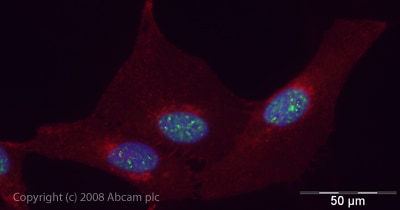 Immunocytochemistry/ Immunofluorescence - Anti-UAP56 antibody (ab47955)ICC/IF image of ab47955 stained human HeLa cells. The cells were PFA fixed (10 min), permabilised in PBS-T (20 min) and incubated with the antibody (ab47955, 1µg/ml) for 1h at room temperature. 1%BSA / 10% normal goat serum / 0.3M glycine was used to quench autofluorescence and block non-specific protein-protein interactions. The secondary antibody (green) was Alexa Fluor® 488 goat anti-rabbit IgG (H+L) used at a 1/1000 dilution for 1h. Alexa Fluor® 594 WGA was used to label plasma membranes (red). DAPI was used to stain the cell nuclei (blue).
 -
 Immunohistochemistry (Formalin/PFA-fixed paraffin-embedded sections) - Anti-UAP56 antibody (ab47955)IHC image of ab47955 staining in human tonsil formalin fixed paraffin embedded tissue section, performed on a Leica BondTM system using the standard protocol F. The section was pre-treated using heat mediated antigen retrieval with sodium citrate buffer (pH6, epitope retrieval solution 1) for 20 mins. The section was then incubated with ab47955, 1µg/ml, for 15 mins at room temperature and detected using an HRP conjugated compact polymer system. DAB was used as the chromogen. The section was then counterstained with haematoxylin and mounted with DPX.
For other IHC staining systems (automated and non-automated) customers should optimize variable parameters such as antigen retrieval conditions, primary antibody concentration and antibody incubation times.
Protocols
Datasheets and documents
References (4)
ab47955 has been referenced in 4 publications.
- Ben-Yishay R et al. Imaging within single NPCs reveals NXF1's role in mRNA export on the cytoplasmic side of the pore. J Cell Biol 218:2962-2981 (2019). PubMed: 31375530
- Galarza-Muñoz G et al. Human Epistatic Interaction Controls IL7R Splicing and Increases Multiple Sclerosis Risk. Cell 169:72-84.e13 (2017). PubMed: 28340352
- Carvalho T et al. Pharmacological inhibition of the spliceosome subunit SF3b triggers exon junction complex-independent nonsense-mediated decay. J Cell Sci 130:1519-1531 (2017). PubMed: 28302904
- Read EK & Digard P Individual influenza A virus mRNAs show differential dependence on cellular NXF1/TAP for their nuclear export. J Gen Virol 91:1290-301 (2010). WB, In situ hybridization ; Human . PubMed: 20071484
Images
-
Western blot - Anti-UAP56 antibody (ab47955)All lanes : Anti-UAP56 antibody (ab47955) at 1 µg/ml
Lane 1 : HeLa (Human epithelial carcinoma cell line) Whole Cell Lysate
Lane 2 : Jurkat (Human T cell lymphoblast-like cell line) Whole Cell Lysate
Lane 3 : HepG2 (Human hepatocellular liver carcinoma cell line) Whole Cell Lysate
Lane 4 : A431 (Human epithelial carcinoma cell line) Whole Cell Lysate
Lysates/proteins at 10 µg per lane.
Secondary
All lanes : IRDye 680 Conjugated Goat Anti-Rabbit IgG (H+L) at 1/10000 dilution
Performed under reducing conditions.
Predicted band size: 49 kDa
Observed band size: 49 kDa
-
Immunocytochemistry/ Immunofluorescence - Anti-UAP56 antibody (ab47955)ICC/IF image of ab47955 stained human HeLa cells. The cells were PFA fixed (10 min), permabilised in PBS-T (20 min) and incubated with the antibody (ab47955, 1µg/ml) for 1h at room temperature. 1%BSA / 10% normal goat serum / 0.3M glycine was used to quench autofluorescence and block non-specific protein-protein interactions. The secondary antibody (green) was Alexa Fluor® 488 goat anti-rabbit IgG (H+L) used at a 1/1000 dilution for 1h. Alexa Fluor® 594 WGA was used to label plasma membranes (red). DAPI was used to stain the cell nuclei (blue).
-
Immunohistochemistry (Formalin/PFA-fixed paraffin-embedded sections) - Anti-UAP56 antibody (ab47955)IHC image of ab47955 staining in human tonsil formalin fixed paraffin embedded tissue section, performed on a Leica BondTM system using the standard protocol F. The section was pre-treated using heat mediated antigen retrieval with sodium citrate buffer (pH6, epitope retrieval solution 1) for 20 mins. The section was then incubated with ab47955, 1µg/ml, for 15 mins at room temperature and detected using an HRP conjugated compact polymer system. DAB was used as the chromogen. The section was then counterstained with haematoxylin and mounted with DPX.
For other IHC staining systems (automated and non-automated) customers should optimize variable parameters such as antigen retrieval conditions, primary antibody concentration and antibody incubation times.


