Anti-U2AF65 antibody (ab37530)
")
Key features and details
- Rabbit polyclonal to U2AF65
- Suitable for: IP, IHC-P, ICC/IF, WB
- Reacts with: Human, Zebrafish
- Isotype: IgG
Overview
-
Product name
Anti-U2AF65 antibody
See all U2AF65 primary antibodies -
Description
Rabbit polyclonal to U2AF65 -
Host species
Rabbit -
Tested applications
Suitable for: IP, IHC-P, ICC/IF, WBmore details -
Species reactivity
Reacts with: Human, Zebrafish
Predicted to work with: Mouse, Cow, Xenopus laevis
-
Immunogen
Synthetic peptide corresponding to Human U2AF65 aa 200-300.
(Peptide available asab37529)
Properties
-
Form
Liquid -
Storage instructions
Shipped at 4°C. Store at +4°C short term (1-2 weeks). Upon delivery aliquot. Store at -20°C or -80°C. Avoid freeze / thaw cycle. -
Storage buffer
pH: 7.40
Preservative: 0.02% Sodium azide
Constituent: PBS
Batches of this product that have a concentration Concentration information loading...
Purity
Immunogen affinity purifiedClonality
PolyclonalIsotype
IgGResearch areas
Associated products
-
Compatible Secondaries
-
Isotype control
Applications
Our Abpromise guarantee covers the use of ab37530 in the following tested applications.
The application notes include recommended starting dilutions; optimal dilutions/concentrations should be determined by the end user.
Application Abreviews Notes IP Use at an assay dependent concentration. IHC-P Use a concentration of 5 µg/ml. ICC/IF Use a concentration of 1 µg/ml. WB 1/250. Detects a band of approximately 65 kDa (predicted molecular weight: 53 kDa). Target
-
Function
Necessary for the splicing of pre-mRNA. Induces cardiac troponin-T (TNNT2) pre-mRNA exon inclusion in muscle. Regulates the TNNT2 exon 5 inclusion through competition with MBNL1. Binds preferentially to a single-stranded structure within the polypyrimidine tract of TNNT2 intron 4 during spliceosome assembly. Required for the export of mRNA out of the nucleus, even if the mRNA is encoded by an intron-less gene. Represses the splicing of MAPT/Tau exon 10. -
Sequence similarities
Belongs to the splicing factor SR family.
Contains 3 RRM (RNA recognition motif) domains. -
Post-translational
modificationsLysyl-hydroxylation at Lys-15 and Lys-276 affects the mRNA splicing activity of the protein, leading to regulate some, but not all, alternative splicing events. -
Cellular localization
Nucleus. - Information by UniProt
-
Database links
- Entrez Gene: 11338 Human
- Entrez Gene: 22185 Mouse
- Entrez Gene: 380287 Xenopus laevis
- Omim: 191318 Human
- SwissProt: P26368 Human
- SwissProt: P26369 Mouse
- Unigene: 528007 Human
- Unigene: 360389 Mouse
-
Alternative names
- hU2AF(65) antibody
- hU2AF65 antibody
- Splicing factor U2AF 65 kDa subunit antibody
see all
Images
-
Western blot - Anti-U2AF65 antibody (ab37530)All lanes : Anti-U2AF65 antibody (ab37530) at 1/250 dilution
Lane 1 : HeLa (Human epithelial carcinoma cell line) Whole Cell Lysate
Lane 2 :Jurkat whole cell lysate (ab7899)
Lane 3 :A-431 whole cell lysate (ab7909)
Lane 4 :HEK-293 whole cell lysate (ab7902)
Lane 5 : Hep G2 whole cell lysate (ab7900)
Lane 6 : MCF-7 (Human breast adenocarcinoma cell line) Whole Cell Lysate
Lane 7 : SHSY-5Y (Human neuroblastoma cell line) Whole Cell Lysate
Lane 8 : U2OS (Human osteosarcoma cell line) Whole Cell Lysate
Lysates/proteins at 10 µg per lane.
Secondary
All lanes : Rabbit IgG secondary antibody (ab28446) at 1/10000 dilution
Performed under reducing conditions.
Predicted band size: 53 kDa
Observed band size: 65 kDa why is the actual band size different from the predicted?Although the predicted band size is 53kDa based on Swiss-prot data, a band of 65kDa has been previously observed. J Biol Chem. 2004 Nov 26;279(48):49773-9 (PMID: 15377657)
-
 Immunocytochemistry/ Immunofluorescence - Anti-U2AF65 antibody (ab37530)ICC/IF image of ab37530 stained human HeLa cells. The cells were PFA fixed (10 min) and incubated with the antibody (ab37530, 1µg/ml) for 1h at room temperature. The secondary antibody (green) was Alexa Fluor® 488 goat anti-rabbit IgG (H+L) used at a 1/1000 dilution for 1h. Image-iTTM FX Signal Enhancer was used to quench autofluorescence. 5% BSA (in TBS-T) was used for all other blocking steps. DAPI was used to stain the cell nuclei (blue). Alexa Fluor® 594 WGA was used to label plasma membranes (red).
 -
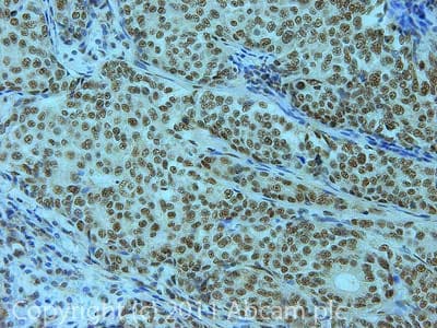 Immunohistochemistry (Formalin/PFA-fixed paraffin-embedded sections) - Anti-U2AF65 antibody (ab37530)IHC image of ab37530 staining in Human breast adenocarcinoma formalin fixed paraffin embedded tissue section, performed on a Leica BondTM system using the standard protocol F. The section was pre-treated using heat mediated antigen retrieval with sodium citrate buffer (pH6, epitope retrieval solution 1) for 20 mins. The section was then incubated with ab37530, 5µg/ml, for 15 mins at room temperature and detected using an HRP conjugated compact polymer system. DAB was used as the chromogen. The section was then counterstained with haematoxylin and mounted with DPX.
For other IHC staining systems (automated and non-automated) customers should optimize variable parameters such as antigen retrieval conditions, primary antibody concentration and antibody incubation times. -
 Western blot - Anti-U2AF65 antibody (ab37530)All lanes : Anti-U2AF65 antibody (ab37530) at 1/250 dilution
Lane 1 : Marker
Lane 2 : Zebrafish brain homogenate at 20 µg
Lane 3 : Zebrafish heart homogenate at 10 µg
Lane 4 : Zebrafish liver homogenate at 10 µg
Lane 5 : Zebrafish skeletal muscle homogenate at 10 µg
Lane 6 : HeLa (Human epithelial carcinoma cell line) Whole Cell Lysate at 10 µg
Secondary
All lanes : Goat polyclonal to Rabbit IgG – H&L – Pre-Adsorbed (HRP) at 1/6000 dilution
Developed using the ECL technique.
Performed under reducing conditions.
Predicted band size: 53 kDa
Observed band size: 53 kDa
Exposure time: 5 minutes -
 Immunoprecipitation - Anti-U2AF65 antibody (ab37530)U2AF65 was immunoprecipitated using 0.5mg SHSY5Y whole cell extract, 5µg of Rabbit polyclonal to U2AF65 and 50µl of protein G magnetic beads (+). No antibody was added to the control (-).
The antibody was incubated under agitation with Protein G beads for 10min, SHSY5Y whole cell extract lysate diluted in RIPA buffer was added to each sample and incubated for a further 10min under agitation.
Proteins were eluted by addition of 40µl SDS loading buffer and incubated for 10min at 70oC; 10µl of each sample was separated on a SDS PAGE gel, transferred to a nitrocellulose membrane, blocked with 5% BSA and probed with ab37530.
Secondary: Mouse monoclonal [SB62a] Secondary Antibody to Rabbit IgG light chain (HRP) (ab99697).
Band: 65kDa: U2AF65.
Protocols
Datasheets and documents
References (13)
ab37530 has been referenced in 13 publications.
- Yuan T et al. Low-density lipoprotein receptor-related protein 6 regulates alternative pre-mRNA splicing. J Cell Mol Med 22:4653-4663 (2018). PubMed: 30070011
- Sithole N et al. DDX17 Specifically, and Independently of DDX5, Controls Use of the HIV A4/5 Splice Acceptor Cluster and Is Essential for Efficient Replication of HIV. J Mol Biol 430:3111-3128 (2018). PubMed: 30131116
- Rodríguez-Mateo C et al. Downregulation of Lnc-Spry1 mediates TGF-ß-induced epithelial-mesenchymal transition by transcriptional and posttranscriptional regulatory mechanisms. Cell Death Differ 24:785-797 (2017). PubMed: 28186499
- Moriel-Carretero M et al. Fanconi anemia FANCD2 and FANCI proteins regulate the nuclear dynamics of splicing factors. J Cell Biol 216:4007-4026 (2017). PubMed: 29030393
- Hou W et al. Two Polypyrimidine Tracts in Intron 4 of the Major Immediate Early Gene Are Critical for Gene Expression Switching from IE1 to IE2 and for Replication of Human Cytomegalovirus. J Virol 90:7339-49 (2016). ChIP ; Human . PubMed: 27252533
Images
-
Western blot - Anti-U2AF65 antibody (ab37530)All lanes : Anti-U2AF65 antibody (ab37530) at 1/250 dilution
Lane 1 : HeLa (Human epithelial carcinoma cell line) Whole Cell Lysate
Lane 2 :Jurkat whole cell lysate (ab7899)
Lane 3 :A-431 whole cell lysate (ab7909)
Lane 4 :HEK-293 whole cell lysate (ab7902)
Lane 5 : Hep G2 whole cell lysate (ab7900)
Lane 6 : MCF-7 (Human breast adenocarcinoma cell line) Whole Cell Lysate
Lane 7 : SHSY-5Y (Human neuroblastoma cell line) Whole Cell Lysate
Lane 8 : U2OS (Human osteosarcoma cell line) Whole Cell Lysate
Lysates/proteins at 10 µg per lane.
Secondary
All lanes : Rabbit IgG secondary antibody (ab28446) at 1/10000 dilution
Performed under reducing conditions.
Predicted band size: 53 kDa
Observed band size: 65 kDa why is the actual band size different from the predicted?Although the predicted band size is 53kDa based on Swiss-prot data, a band of 65kDa has been previously observed. J Biol Chem. 2004 Nov 26;279(48):49773-9 (PMID: 15377657)
-
Immunocytochemistry/ Immunofluorescence - Anti-U2AF65 antibody (ab37530)ICC/IF image of ab37530 stained human HeLa cells. The cells were PFA fixed (10 min) and incubated with the antibody (ab37530, 1µg/ml) for 1h at room temperature. The secondary antibody (green) was Alexa Fluor® 488 goat anti-rabbit IgG (H+L) used at a 1/1000 dilution for 1h. Image-iTTM FX Signal Enhancer was used to quench autofluorescence. 5% BSA (in TBS-T) was used for all other blocking steps. DAPI was used to stain the cell nuclei (blue). Alexa Fluor® 594 WGA was used to label plasma membranes (red).
-
Immunohistochemistry (Formalin/PFA-fixed paraffin-embedded sections) - Anti-U2AF65 antibody (ab37530)IHC image of ab37530 staining in Human breast adenocarcinoma formalin fixed paraffin embedded tissue section, performed on a Leica BondTM system using the standard protocol F. The section was pre-treated using heat mediated antigen retrieval with sodium citrate buffer (pH6, epitope retrieval solution 1) for 20 mins. The section was then incubated with ab37530, 5µg/ml, for 15 mins at room temperature and detected using an HRP conjugated compact polymer system. DAB was used as the chromogen. The section was then counterstained with haematoxylin and mounted with DPX.
For other IHC staining systems (automated and non-automated) customers should optimize variable parameters such as antigen retrieval conditions, primary antibody concentration and antibody incubation times. -
Western blot - Anti-U2AF65 antibody (ab37530)All lanes : Anti-U2AF65 antibody (ab37530) at 1/250 dilution
Lane 1 : Marker
Lane 2 : Zebrafish brain homogenate at 20 µg
Lane 3 : Zebrafish heart homogenate at 10 µg
Lane 4 : Zebrafish liver homogenate at 10 µg
Lane 5 : Zebrafish skeletal muscle homogenate at 10 µg
Lane 6 : HeLa (Human epithelial carcinoma cell line) Whole Cell Lysate at 10 µg
Secondary
All lanes : Goat polyclonal to Rabbit IgG – H&L – Pre-Adsorbed (HRP) at 1/6000 dilution
Developed using the ECL technique.
Performed under reducing conditions.
Predicted band size: 53 kDa
Observed band size: 53 kDa
Exposure time: 5 minutes
-
Immunoprecipitation - Anti-U2AF65 antibody (ab37530)U2AF65 was immunoprecipitated using 0.5mg SHSY5Y whole cell extract, 5µg of Rabbit polyclonal to U2AF65 and 50µl of protein G magnetic beads (+). No antibody was added to the control (-).
The antibody was incubated under agitation with Protein G beads for 10min, SHSY5Y whole cell extract lysate diluted in RIPA buffer was added to each sample and incubated for a further 10min under agitation.
Proteins were eluted by addition of 40µl SDS loading buffer and incubated for 10min at 70oC; 10µl of each sample was separated on a SDS PAGE gel, transferred to a nitrocellulose membrane, blocked with 5% BSA and probed with ab37530.
Secondary: Mouse monoclonal [SB62a] Secondary Antibody to Rabbit IgG light chain (HRP) (ab99697).
Band: 65kDa: U2AF65.


