Anti-Sprouty 1/Spry-1 antibody (ab111523)
Key features and details
- Rabbit polyclonal to Sprouty 1/Spry-1
- Suitable for: ICC/IF, IHC-P, WB
- Reacts with: Human
- Isotype: IgG
Overview
-
Product name
Anti-Sprouty 1/Spry-1 antibody -
Description
Rabbit polyclonal to Sprouty 1/Spry-1 -
Host species
Rabbit -
Tested applications
Suitable for: ICC/IF, IHC-P, WBmore details -
Species reactivity
Reacts with: Human
Predicted to work with: Rabbit, Horse, Cow, Dog, Pig, Macaque monkey, Gorilla, Orangutan
-
Immunogen
Synthetic peptide. This information is proprietary to Abcam and/or its suppliers.
-
Positive control
- WB: Human liver and fetal liver tissue lysates. ICC/IF: methanol fixed HeLa cells. IHC-P: Human normal liver tissue.
-
General notes
The Life Science industry has been in the grips of a reproducibility crisis for a number of years. Abcam is leading the way in addressing this with our range of recombinant monoclonal antibodies and knockout edited cell lines for gold-standard validation. Please check that this product meets your needs before purchasing.
If you have any questions, special requirements or concerns, please send us an inquiry and/or contact our Support team ahead of purchase. Recommended alternatives for this product can be found below, along with publications, customer reviews and Q&As
Properties
-
Form
Liquid -
Storage instructions
Shipped at 4°C. Store at +4°C short term (1-2 weeks). Upon delivery aliquot. Store at -20°C or -80°C. Avoid freeze / thaw cycle. -
Storage buffer
pH: 7.40
Preservative: 0.02% Sodium azide
Constituent: PBS
Batches of this product that have a concentration Concentration information loading...
Purity
Immunogen affinity purifiedClonality
PolyclonalIsotype
IgGResearch areas
Associated products
-
Compatible Secondaries
-
Isotype control
-
Recombinant Protein
Applications
The Abpromise guarantee
Our Abpromise guarantee covers the use of ab111523 in the following tested applications.
The application notes include recommended starting dilutions; optimal dilutions/concentrations should be determined by the end user.
Application Abreviews Notes ICC/IF Use a concentration of 1 µg/ml.IHC-P Use a concentration of 10 µg/ml.WB Use a concentration of 1 µg/ml. Detects a band of approximately 35 kDa (predicted molecular weight: 35 kDa).Notes ICC/IF
Use a concentration of 1 µg/ml.IHC-P
Use a concentration of 10 µg/ml.WB
Use a concentration of 1 µg/ml. Detects a band of approximately 35 kDa (predicted molecular weight: 35 kDa).Target
-
Function
May function as an antagonist of fibroblast growth factor (FGF) pathways and may negatively modulate respiratory organogenesis. -
Sequence similarities
Belongs to the sprouty family.
Contains 1 SPR (sprouty) domain. -
Domain
The Cys-rich domain is responsible for the localization of the protein to the membrane ruffles. -
Cellular localization
Cytoplasm. Membrane. Found in the cytoplasm in unstimulated cells but is translocated to the membrane ruffles in cells stimulated with EGF. - Information by UniProt
-
Database links
- Entrez Gene: 10252 Human
- Omim: 602465 Human
- SwissProt: A5D992 Cow
- SwissProt: O43609 Human
- Unigene: 436944 Human
-
Alternative names
- hSPRY1 antibody
- Protein sprouty homolog 1 antibody
- Sprouty 1 antibody
see all
Images
-
All lanes : Anti-Sprouty 1/Spry-1 antibody (ab111523) at 1 µg/ml
Lane 1 : Human liver tissue lysate - total protein (ab29889)
Lane 2 : Liver (Human) Tissue Lysate - fetal normal tissue (ab29890)
Lysates/proteins at 10 µg per lane.
Secondary
All lanes : Goat Anti-Rabbit IgG H&L (HRP) preadsorbed (ab97080) at 1/5000 dilution
Developed using the ECL technique.
Performed under reducing conditions.
Predicted band size: 35 kDa
Observed band size: 35 kDa
Additional bands at: 85 kDa. We are unsure as to the identity of these extra bands.
Exposure time: 2 minutes -
ICC/IF image of ab111523 stained HeLa cells. The cells were 100% methanol fixed (5 min) and then incubated in 1%BSA / 10% normal goat serum / 0.3M glycine in 0.1% PBS-Tween for 1h to permeabilise the cells and block non-specific protein-protein interactions. The cells were then incubated with the antibody ab111523 at 1µg/ml overnight at +4°C. The secondary antibody (green) was DyLight® 488 goat anti- rabbit (ab96899) IgG (H+L) used at a 1/250 dilution for 1h. Alexa Fluor® 594 WGA was used to label plasma membranes (red) at a 1/200 dilution for 1h. DAPI was used to stain the cell nuclei (blue) at a concentration of 1.43µM.
-
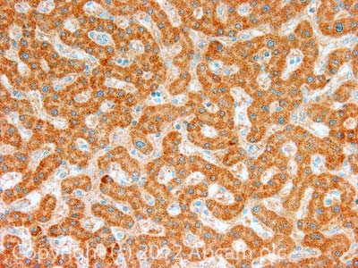
 Immunohistochemistry (Formalin/PFA-fixed paraffin-embedded sections) - Anti-Sprouty 1/Spry-1 antibody (ab111523)
IHC image of Sprouty 1/Spry-1 staining in Human normal liver formalin fixed paraffin embedded tissue section, performed on a Leica BondTM system using the standard protocol F. The section was pre-treated using heat mediated antigen retrieval with EDTA based pH 9.0 solution (epitope retrieval solution 2) for 20 mins. The section was then incubated with ab111523, 10µg/ml, for 15 mins at room temperature and detected using an HRP conjugated compact polymer system. DAB was used as the chromogen. The section was then counterstained with haematoxylin and mounted with DPX.
For other IHC staining systems (automated and non-automated) customers should optimize variable parameters such as antigen retrieval conditions, primary antibody concentration and antibody incubation times
Protocols
Datasheets and documents
-
SDS download
-
Datasheet download
References (4)
ab111523 has been referenced in 4 publications.
- Li Y et al. Injectable hydrogel with MSNs/microRNA-21-5p delivery enables both immunomodification and enhanced angiogenesis for myocardial infarction therapy in pigs. Sci Adv 7:N/A (2021). PubMed: 33627421
- Feng M et al. Circ_0020093 ameliorates IL-1ß-induced apoptosis and extracellular matrix degradation of human chondrocytes by upregulating SPRY1 via targeting miR-23b. Mol Cell Biochem N/A:N/A (2021). PubMed: 34046827
- Qiu B et al. Sprouty4 correlates with favorable prognosis in perihilar cholangiocarcinoma by blocking the FGFR-ERK signaling pathway and arresting the cell cycle. EBioMedicine 50:166-177 (2019). PubMed: 31761616
- Mohis M et al. Aging-related increase in store-operated Ca2+ influx in human ventricular fibroblasts. Am J Physiol Heart Circ Physiol 315:H83-H91 (2018). PubMed: 29985070
Images
-
All lanes : Anti-Sprouty 1/Spry-1 antibody (ab111523) at 1 µg/ml
Lane 1 : Human liver tissue lysate - total protein (ab29889)
Lane 2 : Liver (Human) Tissue Lysate - fetal normal tissue (ab29890)
Lysates/proteins at 10 µg per lane.
Secondary
All lanes : Goat Anti-Rabbit IgG H&L (HRP) preadsorbed (ab97080) at 1/5000 dilution
Developed using the ECL technique.
Performed under reducing conditions.
Predicted band size: 35 kDa
Observed band size: 35 kDa
Additional bands at: 85 kDa. We are unsure as to the identity of these extra bands.
Exposure time: 2 minutes
-
ICC/IF image of ab111523 stained HeLa cells. The cells were 100% methanol fixed (5 min) and then incubated in 1%BSA / 10% normal goat serum / 0.3M glycine in 0.1% PBS-Tween for 1h to permeabilise the cells and block non-specific protein-protein interactions. The cells were then incubated with the antibody ab111523 at 1µg/ml overnight at +4°C. The secondary antibody (green) was DyLight® 488 goat anti- rabbit (ab96899) IgG (H+L) used at a 1/250 dilution for 1h. Alexa Fluor® 594 WGA was used to label plasma membranes (red) at a 1/200 dilution for 1h. DAPI was used to stain the cell nuclei (blue) at a concentration of 1.43µM.
-
 Immunohistochemistry (Formalin/PFA-fixed paraffin-embedded sections) - Anti-Sprouty 1/Spry-1 antibody (ab111523)
IHC image of Sprouty 1/Spry-1 staining in Human normal liver formalin fixed paraffin embedded tissue section, performed on a Leica BondTM system using the standard protocol F. The section was pre-treated using heat mediated antigen retrieval with EDTA based pH 9.0 solution (epitope retrieval solution 2) for 20 mins. The section was then incubated with ab111523, 10µg/ml, for 15 mins at room temperature and detected using an HRP conjugated compact polymer system. DAB was used as the chromogen. The section was then counterstained with haematoxylin and mounted with DPX.
For other IHC staining systems (automated and non-automated) customers should optimize variable parameters such as antigen retrieval conditions, primary antibody concentration and antibody incubation times


