Anti-SHPRH antibody (ab80129)
")
Key features and details
- Rabbit polyclonal to SHPRH
- Suitable for: ICC/IF, WB
- Reacts with: Human
- Isotype: IgG
Overview
-
Product name
Anti-SHPRH antibody
See all SHPRH primary antibodies -
Description
Rabbit polyclonal to SHPRH -
Host species
Rabbit -
Tested Applications & Species
See all applications and species dataApplication Species ICC/IF HumanWB Human
-
Immunogen
Synthetic peptide corresponding to Human SHPRH aa 600-700 conjugated to keyhole limpet haemocyanin.
(Peptide available asab89391) -
Positive control
- This antibody gave a positive signal in SHSY-5Y whole cell lysate.
Properties
-
Form
Liquid -
Storage instructions
Shipped at 4°C. Store at +4°C short term (1-2 weeks). Upon delivery aliquot. Store at -20°C or -80°C. Avoid freeze / thaw cycle. -
Storage buffer
pH: 7.40
Preservative: 0.02% Sodium azide
Constituent: PBS
Batches of this product that have a concentration Concentration information loading...
Purity
Immunogen affinity purifiedClonality
PolyclonalIsotype
IgGResearch areas
Associated products
-
Compatible Secondaries
-
Isotype control
Applications
The Abpromise guarantee
Our Abpromise guarantee covers the use of ab80129 in the following tested applications.
The application notes include recommended starting dilutions; optimal dilutions/concentrations should be determined by the end user.
GuaranteedTested applications are guaranteed to work and covered by our Abpromise guarantee.
PredictedPredicted to work for this combination of applications and species but not guaranteed.
IncompatibleDoes not work for this combination of applications and species.
Application Species ICC/IF HumanWB HumanApplication Abreviews Notes ICC/IF Use a concentration of 5 µg/ml.WB (2) Use a concentration of 1 µg/ml. Detects a band of approximately 193 kDa (predicted molecular weight: 193 kDa).Notes ICC/IF
Use a concentration of 5 µg/ml.WB
Use a concentration of 1 µg/ml. Detects a band of approximately 193 kDa (predicted molecular weight: 193 kDa).Target
-
Function
E3 ubiquitin-protein ligase involved in DNA repair. Upon genotoxic stress, accepts ubiquitin from the UBE2N-UBE2V2 E2 complex and transfers it to 'Lys-164' of PCNA which had been monoubiquitinated by UBE2A/B-RAD18, promoting the formation of non-canonical poly-ubiquitin chains linked through 'Lys-63'. -
Tissue specificity
Broadly expressed. -
Pathway
Protein modification; protein ubiquitination. -
Sequence similarities
Belongs to the SNF2/RAD54 helicase family.
Contains 1 H15 (linker histone H1/H5 globular) domain.
Contains 1 helicase ATP-binding domain.
Contains 1 helicase C-terminal domain.
Contains 1 PHD-type zinc finger.
Contains 1 RING-type zinc finger. -
Domain
The RING finger mediates E3 ubiquitin ligase activity. - Information by UniProt
-
Database links
- Entrez Gene: 257218 Human
- Omim: 608048 Human
- SwissProt: Q149N8 Human
- Unigene: 723297 Human
-
Alternative names
- bA545I5.2 antibody
- E3 ubiquitin-protein ligase SHPRH antibody
- FLJ27258 antibody
see all
Images
-
Western blot - Anti-SHPRH antibody (ab80129)This image is courtesy of an anonymous AbreviewAnti-SHPRH antibody (ab80129) at 1/1000 dilution + HEK293 whole cell lysate at 50 µg
Secondary
HRP-conjugated goat anti-rabbit IgG polyclonal at 1/10000 dilution
Developed using the ECL technique.
Performed under reducing conditions.
Predicted band size: 193 kDa
Observed band size: 175 kDa why is the actual band size different from the predicted?
Exposure time: 5 minutes
-
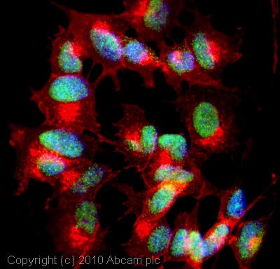 Immunocytochemistry/ Immunofluorescence - Anti-SHPRH antibody (ab80129)ICC/IF image of ab80129 stained SHSY5Y cells. The cells were 4% formaldehyde fixed (10 min) and then incubated in 1%BSA / 10% normal goat serum / 0.3M glycine in 0.1% PBS-Tween for 1h to permeabilise the cells and block non-specific protein-protein interactions. The cells were then incubated with the antibody (ab80129, 5µg/ml) overnight at +4°C. The secondary antibody (green) was Alexa Fluor® 488 goat anti-rabbit IgG (H+L) used at a 1/1000 dilution for 1h. Alexa Fluor® 594 WGA was used to label plasma membranes (red) at a 1/200 dilution for 1h. DAPI was used to stain the cell nuclei (blue) at a concentration of 1.43µM. This antibody also gave a positive result in 100% methanol fixed (5 min) SHSY5Y cells at 5µg/ml.
 -
 Western blot - Anti-SHPRH antibody (ab80129)Anti-SHPRH antibody (ab80129) at 1 µg/ml + SHSY-5Y (Human neuroblastoma cell line) Whole Cell Lysate at 10 µg
Secondary
Goat polyclonal to Rabbit IgG - H&L - Pre-Adsorbed (HRP) at 1/3000 dilution
Developed using the ECL technique.
Performed under reducing conditions.
Predicted band size: 193 kDa
Observed band size: 104,193 kDa why is the actual band size different from the predicted?
Additional bands at: 172 kDa. We are unsure as to the identity of these extra bands.
Exposure time: 5 minutes
This antibody was raised against an immunogen that is predicted to recognize isoforms (1,2,3,4 and 5) of E3 ubiquitin-protein ligase SHPRH. The predicted molecular weights of isoforms (1,2,3,4 and 5) are 193 kDa, 104kDa, 17 kDa, 190 kDa and 122 kDa respectively.
Protocols
Datasheets and documents
References (2)
ab80129 has been referenced in 2 publications.
- Waraky A et al. Nuclear insulin-like growth factor 1 receptor phosphorylates proliferating cell nuclear antigen and rescues stalled replication forks after DNA damage. J Biol Chem 292:18227-18239 (2017). PubMed: 28924044
- Qu Y et al. Axitinib blocks Wnt/ß-catenin signaling and directs asymmetric cell division in cancer. Proc Natl Acad Sci U S A 113:9339-44 (2016). WB . PubMed: 27482107
Images
-
Western blot - Anti-SHPRH antibody (ab80129) This image is courtesy of an anonymous AbreviewAnti-SHPRH antibody (ab80129) at 1/1000 dilution + HEK293 whole cell lysate at 50 µg
Secondary
HRP-conjugated goat anti-rabbit IgG polyclonal at 1/10000 dilution
Developed using the ECL technique.
Performed under reducing conditions.
Predicted band size: 193 kDa
Observed band size: 175 kDa why is the actual band size different from the predicted?
Exposure time: 5 minutes
-
Immunocytochemistry/ Immunofluorescence - Anti-SHPRH antibody (ab80129)ICC/IF image of ab80129 stained SHSY5Y cells. The cells were 4% formaldehyde fixed (10 min) and then incubated in 1%BSA / 10% normal goat serum / 0.3M glycine in 0.1% PBS-Tween for 1h to permeabilise the cells and block non-specific protein-protein interactions. The cells were then incubated with the antibody (ab80129, 5µg/ml) overnight at +4°C. The secondary antibody (green) was Alexa Fluor® 488 goat anti-rabbit IgG (H+L) used at a 1/1000 dilution for 1h. Alexa Fluor® 594 WGA was used to label plasma membranes (red) at a 1/200 dilution for 1h. DAPI was used to stain the cell nuclei (blue) at a concentration of 1.43µM. This antibody also gave a positive result in 100% methanol fixed (5 min) SHSY5Y cells at 5µg/ml.
-
Western blot - Anti-SHPRH antibody (ab80129)Anti-SHPRH antibody (ab80129) at 1 µg/ml + SHSY-5Y (Human neuroblastoma cell line) Whole Cell Lysate at 10 µg
Secondary
Goat polyclonal to Rabbit IgG - H&L - Pre-Adsorbed (HRP) at 1/3000 dilution
Developed using the ECL technique.
Performed under reducing conditions.
Predicted band size: 193 kDa
Observed band size: 104,193 kDa why is the actual band size different from the predicted?
Additional bands at: 172 kDa. We are unsure as to the identity of these extra bands.
Exposure time: 5 minutes
This antibody was raised against an immunogen that is predicted to recognize isoforms (1,2,3,4 and 5) of E3 ubiquitin-protein ligase SHPRH. The predicted molecular weights of isoforms (1,2,3,4 and 5) are 193 kDa, 104kDa, 17 kDa, 190 kDa and 122 kDa respectively.
