Anti-Securin antibody (ab26273)
Key features and details
- Rabbit polyclonal to Securin
- Suitable for: WB, ICC/IF
- Knockout validated
- Reacts with: Human
- Isotype: IgG
Overview
-
Product name
Anti-Securin antibody
See all Securin primary antibodies -
Description
Rabbit polyclonal to Securin -
Host species
Rabbit -
Tested Applications & Species
See all applications and species dataApplication Species ICC/IF HumanWB Human -
Immunogen
Synthetic peptide conjugated to KLH derived from within residues 100 - 200 of Human Securin.
Read Abcam's proprietary immunogen policy -
Positive control
- WB: Wild-type HEK293T cell lysate. Daudi and HEK293 cell lysates.
-
General notes
The Life Science industry has been in the grips of a reproducibility crisis for a number of years. Abcam is leading the way in addressing the problem with our range of recombinant monoclonal antibodies and knockout edited cell lines for gold-standard validation.
One factor contributing to the crisis is the use of antibodies that are not suitable. This can lead to misleading results and the use of incorrect data informing project assumptions and direction. To help address this challenge, we have introduced an application and species grid on our primary antibody datasheets to make it easy to simplify identification of the right antibody for your needs.
Learn more here.
Properties
-
Form
Liquid -
Storage instructions
Shipped at 4°C. Store at +4°C short term (1-2 weeks). Upon delivery aliquot. Store at -20°C or -80°C. Avoid freeze / thaw cycle. -
Storage buffer
pH: 7.4
Preservative: 0.02% Sodium azide
Constituent: PBS
Batches of this product that have a concentration Concentration information loading...
Purity
Immunogen affinity purifiedClonality
PolyclonalIsotype
IgGResearch areas
Associated products
-
Compatible Secondaries
-
Isotype control
-
KO cell lines
-
KO cell lysates
-
Positive Controls
-
Recombinant Protein
Applications
The Abpromise guarantee
Our Abpromise guarantee covers the use of ab26273 in the following tested applications.
The application notes include recommended starting dilutions; optimal dilutions/concentrations should be determined by the end user.
GuaranteedTested applications are guaranteed to work and covered by our Abpromise guarantee.
PredictedPredicted to work for this combination of applications and species but not guaranteed.
IncompatibleDoes not work for this combination of applications and species.
Application Species ICC/IF HumanWB HumanAll applications MouseRatApplication Abreviews Notes WB (4) Use at an assay dependent concentration. Detects a band of approximately 26 kDa (predicted molecular weight: 22 kDa).ICC/IF (1) 1/100.Notes WB
Use at an assay dependent concentration. Detects a band of approximately 26 kDa (predicted molecular weight: 22 kDa).ICC/IF
1/100.Target
-
Function
Regulatory protein, which plays a central role in chromosome stability, in the p53/TP53 pathway, and DNA repair. Probably acts by blocking the action of key proteins. During the mitosis, it blocks Separase/ESPL1 function, preventing the proteolysis of the cohesin complex and the subsequent segregation of the chromosomes. At the onset of anaphase, it is ubiquitinated, conducting to its destruction and to the liberation of ESPL1. Its function is however not limited to a blocking activity, since it is required to activate ESPL1. Negatively regulates the transcriptional activity and related apoptosis activity of TP53. The negative regulation of TP53 may explain the strong transforming capability of the protein when it is overexpressed. May also play a role in DNA repair via its interaction with Ku, possibly by connecting DNA damage-response pathways with sister chromatid separation. -
Tissue specificity
Expressed at low level in most tissues, except in adult testis, where it is highly expressed. Overexpressed in many patients suffering from pituitary adenomas, primary epithelial neoplasias, and esophageal cancer. -
Sequence similarities
Belongs to the securin family. -
Developmental stage
Low level during G1 and S phases. Peaks at M phase. During anaphase, it is degraded. -
Domain
The N-terminal destruction box (D-box) acts as a recognition signal for degradation via the ubiquitin-proteasome pathway.
The TEK-boxes are required for 'Lys-11'-linked ubiquitination and facilitate the transfer of the first ubiquitin and ubiquitin chain nucleation. TEK-boxes may direct a catalytically competent orientation of the UBE2C/UBCH10-ubiquitin thiolester with the acceptor lysine residue. -
Post-translational
modificationsPhosphorylated at Ser-165 by CDK1 during mitosis.
Phosphorylated in vitro by ds-DNA kinase.
Ubiquitinated through 'Lys-11' linkage of ubiquitin moieties by the anaphase promoting complex (APC) at the onset of anaphase, conducting to its degradation. 'Lys-11'-linked ubiquitination is mediated by the E2 ligase UBE2C/UBCH10. -
Cellular localization
Cytoplasm. Nucleus. - Information by UniProt
-
Database links
- Entrez Gene: 9232 Human
- Entrez Gene: 30939 Mouse
- Entrez Gene: 64193 Rat
- Omim: 604147 Human
- SwissProt: O95997 Human
- SwissProt: Q6IAL9 Human
- SwissProt: Q3Y5K4 Mouse
- SwissProt: Q5SRU0 Mouse
see all -
Alternative names
- AW555095 antibody
- C87862 antibody
- Cut2 antibody
see all
Images
-
All lanes : Anti-Securin antibody (ab26273) at 1/500 dilution
Lane 1 : Wild-type HEK293T cell lysate
Lane 2 : PTTG1 knockout HEK293T cell lysate
Lane 3 : Daudi cell lysate
Lysates/proteins at 20 µg per lane.
Performed under reducing conditions.
Predicted band size: 22 kDa
Observed band size: 28 kDa why is the actual band size different from the predicted?Lanes 1-3: Merged signal (red and green). Green - ab26273 observed at 28 kDa. Red - loading control, ab8245 observed at 37 kDa.
ab26273 Anti-Securin antibody was shown to specifically react with Securin in wild-type HEK293T cells. Loss of signal was observed when knockout cell line ab266231 (knockout cell lysate ab257289) was used. Wild-type and Securin knockout samples were subjected to SDS-PAGE. ab26273 and Anti-GAPDH antibody [6C5] - Loading Control (ab8245) were incubated overnight at 4°C at 1 in 500 dilution and 1 in 20000 dilution respectively. Blots were developed with Goat anti-Rabbit IgG H&L (IRDye® 800CW) preadsorbed (ab216773) and Goat anti-Mouse IgG H&L (IRDye® 680RD) preadsorbed (ab216776) secondary antibodies at 1 in 10000 dilution for 1 hour at room temperature before imaging.
-
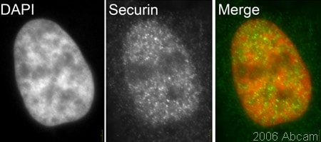
 Immunocytochemistry/ Immunofluorescence - Anti-Securin antibody (ab26273)This image is courtesy of Kirk McManusHeLa cells stained with ab26273 (1/100 dilution). A clear nuclear signal in interphase HeLa cells can be seen.
 -
 Western blot - Anti-Securin antibody (ab26273)This image is courtesy of an abreview submitted by Andrew HorwitzAll lanes : Anti-Securin antibody (ab26273) at 1/500 dilution
Lane 1 : HEK293 human cell lysate
Lane 2 : MPRO mouse cell lysate
Lane 3 : S. Cerevisiae lysate
Lysates/proteins at 50 µg per lane.
Secondary
All lanes : Polyclonal Goat anti Rabbit at 1/10000 dilution
Predicted band size: 22 kDa
-
Anti-Securin antibody (ab26273) at 1 µg/ml + HEK293 (Human embryonic kidney cell line) Whole Cell Lysate at 20 µg
Secondary
Goat polyclonal to Rabbit IgG - H&L - Pre-Adsorbed (HRP) at 1/3000 dilution
Performed under reducing conditions.
Predicted band size: 22 kDa
Observed band size: 26 kDa why is the actual band size different from the predicted?
Exposure time: 15 minutes
ab26273 detects a clean 26 kDa band in HEK293 lysate. We would recommend loading 20-40ug of protein when using this antibody. ab26273 has also been tested in mouse lysates and in our hands detects a 70 kDa band which we cannot explain.
Protocols
References (3)
ab26273 has been referenced in 3 publications.
- Lin X et al. PTTG1 is involved in TNF-a-related hepatocellular carcinoma via the induction of c-myc. Cancer Med 8:5702-5715 (2019). PubMed: 31385458
- Lee SB et al. Parkin Regulates Mitosis and Genomic Stability through Cdc20/Cdh1. Mol Cell 60:21-34 (2015). IP . PubMed: 26387737
- Lubka-Pathak M et al. Altered expression of securin (Pttg1) and serpina3n in the auditory system of hearing-impaired Tff3-deficient mice. Cell Mol Life Sci 68:2739-49 (2011). PubMed: 21076990
Images
-
All lanes : Anti-Securin antibody (ab26273) at 1/500 dilution
Lane 1 : Wild-type HEK293T cell lysate
Lane 2 : PTTG1 knockout HEK293T cell lysate
Lane 3 : Daudi cell lysate
Lysates/proteins at 20 µg per lane.
Performed under reducing conditions.
Predicted band size: 22 kDa
Observed band size: 28 kDa why is the actual band size different from the predicted?Lanes 1-3: Merged signal (red and green). Green - ab26273 observed at 28 kDa. Red - loading control, ab8245 observed at 37 kDa.
ab26273 Anti-Securin antibody was shown to specifically react with Securin in wild-type HEK293T cells. Loss of signal was observed when knockout cell line ab266231 (knockout cell lysate ab257289) was used. Wild-type and Securin knockout samples were subjected to SDS-PAGE. ab26273 and Anti-GAPDH antibody [6C5] - Loading Control (ab8245) were incubated overnight at 4°C at 1 in 500 dilution and 1 in 20000 dilution respectively. Blots were developed with Goat anti-Rabbit IgG H&L (IRDye® 800CW) preadsorbed (ab216773) and Goat anti-Mouse IgG H&L (IRDye® 680RD) preadsorbed (ab216776) secondary antibodies at 1 in 10000 dilution for 1 hour at room temperature before imaging.
-
 Immunocytochemistry/ Immunofluorescence - Anti-Securin antibody (ab26273) This image is courtesy of Kirk McManusHeLa cells stained with ab26273 (1/100 dilution). A clear nuclear signal in interphase HeLa cells can be seen.
 -
 Western blot - Anti-Securin antibody (ab26273) This image is courtesy of an abreview submitted by Andrew HorwitzAll lanes : Anti-Securin antibody (ab26273) at 1/500 dilution
Lane 1 : HEK293 human cell lysate
Lane 2 : MPRO mouse cell lysate
Lane 3 : S. Cerevisiae lysate
Lysates/proteins at 50 µg per lane.
Secondary
All lanes : Polyclonal Goat anti Rabbit at 1/10000 dilution
Predicted band size: 22 kDa
-
Anti-Securin antibody (ab26273) at 1 µg/ml + HEK293 (Human embryonic kidney cell line) Whole Cell Lysate at 20 µg
Secondary
Goat polyclonal to Rabbit IgG - H&L - Pre-Adsorbed (HRP) at 1/3000 dilution
Performed under reducing conditions.
Predicted band size: 22 kDa
Observed band size: 26 kDa why is the actual band size different from the predicted?
Exposure time: 15 minutes
ab26273 detects a clean 26 kDa band in HEK293 lysate. We would recommend loading 20-40ug of protein when using this antibody. ab26273 has also been tested in mouse lysates and in our hands detects a 70 kDa band which we cannot explain.


