Anti-PKC alpha (phospho S657 + Y658) antibody (ab23513)
Key features and details
- Rabbit polyclonal to PKC alpha (phospho S657 + Y658)
- Suitable for: ICC, WB
- Reacts with: Rat
- Isotype: IgG
Overview
-
Product name
Anti-PKC alpha (phospho S657 + Y658) antibody
See all PKC alpha primary antibodies -
Description
Rabbit polyclonal to PKC alpha (phospho S657 + Y658) -
Host species
Rabbit -
Specificity
ab23513 detected PKC alpha and PKC beta I, but not other PKC isoforms in rat brain lysate. -
Tested applications
Suitable for: ICC, WBmore details -
Species reactivity
Reacts with: Rat -
Immunogen
Phospho-PKC alpha (Ser-657/Tyr-658) synthetic peptide (coupled to carrier protein) corresponding to amino acid residues around serine 657 and tyrosine 658 of human PKC alpha.
-
General notes
We do not have concentration information for this antibody. It is optimized to work at the dilutions stated on the datasheet.
Properties
-
Form
Liquid -
Storage instructions
Shipped at 4°C. Store at +4°C short term (1-2 weeks). Upon delivery aliquot. Store at -20°C. Avoid freeze / thaw cycle. -
Storage buffer
Preservative: 0.05% Sodium azide
Constituents: PBS, 50% Glycerol, 0.1% BSA -
 Concentration information loading...
 -
Purity
Affinity purified -
Clonality
Polyclonal -
Isotype
IgG -
Research areas
Images
-
Western blot analysis of immunoprecipitates from neonatal rat brain lysate using anti-PKCα antibody. Control and alkaline phosphatase treated precipitates were probed with anti-PKCα (Central region) or anti-phospho-PKCα (Ser-657/Tyr-658).
-
Immunocytochemical labeling of PKC phosphorylation in aldehyde-fixed and NP-40-permeabilized NGF-differentiated PC12 cells. The cells were labeled with rabbit polyclonal anti-PKCα (Ser-657/Tyr-658) (PP1091) antibody in the absence (Left) or presence (Right) of blocking peptide (PX1095). The antibody was detected using appropriate secondary antibody conjugated to DyLight® 594
-
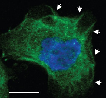
 Immunocytochemistry - Anti-PKC alpha (phospho S657 + Y658) antibody (ab23513) Image from Taniuchi K et al., PLoS One. 2012;7(4):e35674. Epub 2012 Apr 19. Fig 8.; doi:10.1371/journal.pone.0035674; April 19, 2012, PLoS ONE 7(4): e35674.Immunofluorescence analysis of S2-013 cells, staining PKC alpha (phospho S657 + Y658) with ab23513.
Cells were fixed with paraformaldehyde, permeabilized with 0.1% Triton X-100, and blocked with blocking solution (3% BSA/PBS). Cells were incubated with primary antibody for 1 hour. An AlexaFluor®488-conjugated anti-rabbit IgG was used as the secondary antibody.


