Anti-PARP1 antibody (ab227244)
")
Key features and details
- Rabbit polyclonal to PARP1
- Suitable for: ChIP, IHC-P, WB, ICC/IF, IP
- Reacts with: Mouse, Rat, Human
- Isotype: IgG
Overview
-
Product name
Anti-PARP1 antibody
See all PARP1 primary antibodies -
Description
Rabbit polyclonal to PARP1 -
Host species
Rabbit -
Tested Applications & Species
See all applications and species dataApplication Species ChIP HumanICC/IF HumanIHC-P HumanIP HumanWB MouseRatHuman
-
Immunogen
Recombinant fragment within Human PARP1 (internal sequence). The exact sequence is proprietary.
Database link: P09874 -
Positive control
- WB: HEK-293T, NIH/3T3 and PC-12 whole cell extracts; HCT 116, HEK-293T, A431, HeLa and HepG2 whole cell lysate (ab7900). ICC/IF: HeLa cells. ChIP: HeLa and Raji chromatin extracts. IP: HCT 116 whole cell extract. IHC-P: HeLa xenograft tissue.
Properties
-
Form
Liquid -
Storage instructions
Shipped at 4°C. Store at +4°C short term (1-2 weeks). Upon delivery aliquot. Store at -20°C long term. Avoid freeze / thaw cycle. -
Storage buffer
pH: 7.00
Preservative: 0.025% Proclin 300
Constituents: 78% PBS, 1% BSA, 20% Glycerol (glycerin, glycerine) -
 Concentration information loading...
 -
Purity
Immunogen affinity purified -
Clonality
Polyclonal -
Isotype
IgG -
Research areas
Images
-
Western blot - Anti-PARP1 antibody (ab227244)All lanes : Anti-PARP1 antibody (ab227244) at 1/5000 dilution
Lane 1 : Non-transfected HEK-293T (human epithelial cell line from embryonic kidney transformed with large T antigen) whole cell extract
Lane 2 : PARP1 shRNA transfected HEK-293T (human epithelial cell line from embryonic kidney transformed with large T antigen) whole cell extract
Lysates/proteins at 30 µg per lane.
Secondary
All lanes : HRP-conjugated anti-rabbit IgG
Predicted band size: 113 kDa7.5% SDS-PAGE gel.
-
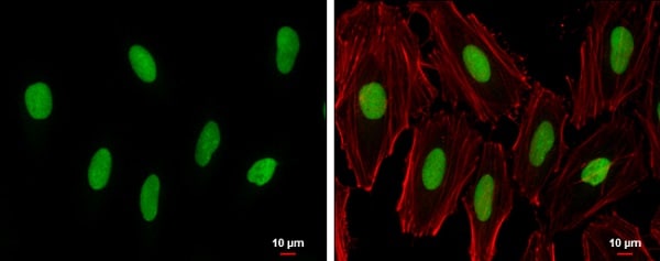 Immunocytochemistry/ Immunofluorescence - Anti-PARP1 antibody (ab227244)
4% paraformaldehyde-fixed HeLa (human epithelial cell line from cervix adenocarcinoma) cells stained for PARP1 (green) using ab227244 at 1/500 dilution in ICC/IF.
Red: Phalloidin, a cytoskeleton marker, at 1/200 dilution.
Blue: Hoechst 33342 staining.
-
 ChIP - Anti-PARP1 antibody (ab227244)
ChIP was performed with HeLa chromatin extract and 5 µg of either normal rabbit IgG or ab227244. The precipitated DNA was detected by PCR with primer set targeting to HSP70.1 promoter.
-
 Immunohistochemistry (Formalin/PFA-fixed paraffin-embedded sections) - Anti-PARP1 antibody (ab227244)
Paraffin-embedded HeLa xenograft tissue stained for PARP1 using ab227244 at 1/500 dilution in immunohistochemical analysis.
Antigen Retrieval: EDTA based, pH 8.0, buffer, 15minutes.
-
 Immunoprecipitation - Anti-PARP1 antibody (ab227244)
PARP1 was immunoprecipitated from HCT 116 (human colorectal carcinoma cell line) whole cell extract with 4 µg ab227244. Western blot was performed from the immunoprecipitate using ab227244 at 1/500 dilution. Anti-Rabbit IgG was used as a secondary reagent.
Lane 1: HCT 116 whole cell extract 30 μg.
Lane 2: Control IP in HCT 116 whole cell extract with 4 μg of preimmune Rabbit IgG.
Lane 3: ab227244 IP in HCT 116 whole cell extract.
-
 Western blot - Anti-PARP1 antibody (ab227244)All lanes : Anti-PARP1 antibody (ab227244) at 1/1000 dilution
Lane 1 : 293T whole cell extracts
Lane 2 : A431 whole cell extracts
Lane 3 : HeLa whole cell extracts
Lane 4 : HepG2 whole cell extracts
Lysates/proteins at 30 µg per lane.
Secondary
All lanes : Rabbit IgG antibody (HRP) at 1/10000 dilution
Predicted band size: 113 kDa5% gel.
Running conditions: 80V, 15min; 140V, 40 minutes.
Transfer condition: Semi-dry, 18 V, 60 min (NC membrane).
Blocking condition: 5% non-fat milk in TBST, RT, 60 minutes.
Primary antibody incubation: 4? , overnight.
Washing condition: 5 ml TBST, 4 x 5 minutes.
Exposure: chemiluminescent substrate for the detection of HRP-conjugated antibody
-
 Western blot - Anti-PARP1 antibody (ab227244)All lanes : Anti-PARP1 antibody (ab227244) at 1/1000 dilution
All lanes : Neuro2A whole cell extracts
Lysates/proteins at 30 µg per lane.
Secondary
All lanes : Rabbit IgG antibody (HRP) at 1/10000 dilution
Predicted band size: 113 kDa5% gel.
Running conditions: 80V, 15min; 140V, 40 minutes.
Transfer condition: Semi-dry, 18 V, 60 min (NC membrane).
Blocking condition: 5% non-fat milk in TBST, RT, 60 minutes.
Primary antibody incubation: 4? , overnight.
Washing condition: 5 ml TBST, 4 x 5 minutes.
Exposure: chemiluminescent substrate for the detection of HRP-conjugated antibody
-
 Western blot - Anti-PARP1 antibody (ab227244)All lanes : Anti-PARP1 antibody (ab227244) at 1/5000 dilution
Lane 1 : HCT116 whole cell lysate (untreated)
Lane 2 : HCT116 whole cell lysate (30 µM cisplatin treatment for 24 hours)
Lysates/proteins at 30 µg per lane.
Secondary
All lanes : HRP-conjugated anti-rabbit IgG
Predicted band size: 113 kDa7.5% SDS-PAGE gel.
The top band is full-length and the bottom band on lane 2 is cleaved PARP1.
-
 ChIP - Anti-PARP1 antibody (ab227244)
Cross-linked ChIP was performed with Raji chromatin extract and 5 μg of either control rabbit IgG or ab227244. The precipitated DNA was detected by PCR with primer set targeting to S100A9 promoter.
-
 Western blot - Anti-PARP1 antibody (ab227244)All lanes : Anti-PARP1 antibody (ab227244) at 1/500 dilution
Lane 1 : NIH/3T3 (mouse embryo fibroblast cell line) whole cell extract
Lane 2 : PC-12 (rat adrenal gland pheochromocytoma cell line) whole cell extract
Secondary
All lanes : HRP-conjugated anti-rabbit IgG
Predicted band size: 113 kDa7.5% SDS-PAGE gel.


