Anti-non-muscle Myosin IIA antibody [2B3] (ab55456)
Key features and details
- Mouse monoclonal [2B3] to non-muscle Myosin IIA
- Suitable for: WB, IHC-P, Flow Cyt, ICC/IF
- Knockout validated
- Reacts with: Human, Recombinant fragment
- Isotype: IgG2b
Overview
-
Product name
Anti-non-muscle Myosin IIA antibody [2B3]
See all non-muscle Myosin IIA primary antibodies -
Description
Mouse monoclonal [2B3] to non-muscle Myosin IIA -
Host species
Mouse -
Tested Applications & Species
See all applications and species dataApplication Species Flow Cyt HumanICC/IF HumanIHC-P HumanWB HumanRecombinant fragment -
Immunogen
Recombinant fragment (GST-tag) corresponding to Human non-muscle Myosin IIA aa 1871-1960. MW of the GST tag alone is 26 KDa.
Sequence:RLKQLKRQLEEAEEEAQRANASRRKLQRELEDATETADAMNREVSSLKNK LRRGDLPFVVPRRMARKGAGDGSDEEVDGKADGAEAKPAE
Database link: P35579 -
Positive control
- WB: HAP1, HeLa ,HEK293 cell lysate; human kidney tissue lysate. ICC/IF: HeLa, MCF7 cells. IHC-P: Human kidney tissue. Flow cyt: HEK293 cells.
-
General notes
Abcam is committed to meeting high standards of ethical manufacturing and has decided to discontinue this product by 01st December 2020 as it has been generated by the ascites method. We recommend ab138498 as an alternative product. We are sorry for any inconvenience this may cause.
Properties
-
Form
Liquid -
Storage instructions
Shipped at 4°C. Store at +4°C short term (1-2 weeks). Upon delivery aliquot. Store at -20°C long term. Avoid freeze / thaw cycle. -
Storage buffer
Constituent: PBS -
 Concentration information loading...
 -
Purity
Protein A purified -
Clonality
Monoclonal -
Clone number
2B3 -
Isotype
IgG2b -
Light chain type
kappa -
Research areas
Images
-
Lane 1: Wild-type HAP1 cell lysate (20 µg)
Lane 2: non-muscle Myosin IIA knockout HAP1 cell lysate (20 µg)
Lane 3: HeLa cell lysate (20 µg)
Lane 4: HEK293 cell lysate (20 µg)
Lanes 1 - 4: Merged signal (red and green). Green - ab55456 observed at 230 kDa. Red - loading control, ab18251, observed at 52 kDa.
ab55456 was shown to specifically react with non-muscle Myosin IIA when non-muscle Myosin IIA knockout samples were used. Wild-type and non-muscle Myosin IIA knockout samples were subjected to SDS-PAGE. ab55456 at a concentration of 1 µg/ml and ab18251 (loading control to alpha Tubulin) at a dilution of 1/1000 were incubated overnight at 4°C. Blots were developed withGoat anti-Mouse IgG H&L (IRDye® 800CW) preadsorbed (ab216772) and Goat Anti-Rabbit IgG H&L (IRDye® 680RD) preadsorbed (ab216777) secondary antibodies at 1/10000 dilution for 1 hour at room temperature before imaging. -
Immunofluorecence analysis of HeLa (human epithelial cell line from cervix adenocarcinoma) cells labeling non-muscle Myosin IIA (Green) using ab55456 at 10 µg/ml in ICC/IF analysis.
-
![Immunocytochemistry/ Immunofluorescence - Anti-non-muscle Myosin IIA antibody [2B3] (ab55456)](https://www.abcam.com/ps/products/55/ab55456/Images/non-muscle-Myosin-IIA-Primary-antibodies-ab55456-9.jpg) Immunocytochemistry/ Immunofluorescence - Anti-non-muscle Myosin IIA antibody [2B3] (ab55456) Image from Bailey CK et al., J Biol Chem. 2012 Jun 1;287(23):19472-86. Epub 2012 Apr 11. Fig 10.; doi: 10.1074/jbc.M112.345728; June 1, 2012 The Journal of Biological Chemistry, 287, 19472-19486.Immunofluorescence analysis of plakoglobin-knocked down MCF7 cells, staining non-muscle Myosin IIA with ab55456.
Immunocytochemistry/ Immunofluorescence - Anti-non-muscle Myosin IIA antibody [2B3] (ab55456) Image from Bailey CK et al., J Biol Chem. 2012 Jun 1;287(23):19472-86. Epub 2012 Apr 11. Fig 10.; doi: 10.1074/jbc.M112.345728; June 1, 2012 The Journal of Biological Chemistry, 287, 19472-19486.Immunofluorescence analysis of plakoglobin-knocked down MCF7 cells, staining non-muscle Myosin IIA with ab55456.
Cells were fixed with formaldehyde, permeabilized with 0.2% Triton X-100 and blocked in 5% goat serum. Samples were incubated with primary antibody overnight at 4°C. An AlexaFluor®-conjugated anti-mouse IgG was used as the secondary antibody. -
 Immunohistochemistry (Formalin/PFA-fixed paraffin-embedded sections) - Anti-non-muscle Myosin IIA antibody (ab55456)
Immunohistochemical analysis of paraffin-embedded human kidney tissue labeling non-muscle Myosin llA with ab55456 at 0.5 µg/ml.
-
Overlay histogram showing HEK293 cells stained with ab55456 (red line). The cells were fixed with 4% paraformaldehyde (10 min) and then permeabilized with 0.1% PBS-Tween for 20 min. The cells were then incubated in 1x PBS / 10% normal goat serum / 0.3M glycine to block non-specific protein-protein interactions followed by the antibody (ab55456, 1µg/1x106 cells) for 30 min at 22°C. The secondary antibody used was DyLight® 488 goat anti-mouse IgG (H+L) (ab96879) at 1/500 dilution for 30 min at 22°C. Isotype control antibody (black line) was mouse IgG2b [PLPV219] (ab91366, 2µg/1x106 cells) used under the same conditions. Acquisition of >5,000 events was performed. This antibody gave a positive signal in HEK293 cells fixed with 100% methanol (5 min)/permeabilized in 0.1% PBS-Tween used under the same conditions.
-
ICC/IF image of ab55456 stained HeLa cells. The cells were 100% methanol fixed (5 min) and then incubated in 1%BSA / 10% normal goat serum / 0.3M glycine in 0.1% PBS-Tween for 1h to permeabilise the cells and block non-specific protein-protein interactions. The cells were then incubated with the antibody (ab55456, 1µg/ml) overnight at +4°C. The secondary antibody (green) was Alexa Fluor® 488 goat anti-mouse IgG (H+L) used at a 1/1000 dilution for 1h. Alexa Fluor® 594 WGA was used to label plasma membranes (red) at a 1/200 dilution for 1h. DAPI was used to stain the cell nuclei (blue) at a concentration of 1.43µM.
-
Western blot against tagged recombinant protein immunogen using ab55456 non-muscle Myosin IIA antibody at 1ug/ml. Predicted band size of immunogen is 33 kDa
-
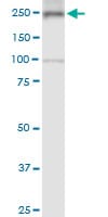
Anti-non-muscle Myosin IIA antibody [2B3] (ab55456) + HeLa
-
Anti-non-muscle Myosin IIA antibody [2B3] (ab55456) at 5 µg/ml + Human kidney tissue lysate
![Anti-non-muscle Myosin IIA antibody [2B3] (ab55456) Anti-non-muscle Myosin IIA antibody [2B3] (ab55456)](https://www.abcam.com/ps/products/55/ab55456/Images/ab55456-260296-ab55456KOWB.jpg)


![Immunocytochemistry/ Immunofluorescence - Anti-non-muscle Myosin IIA antibody [2B3] (ab55456)](https://www.abcam.com/ps/products/55/ab55456/Images/ab55456-355403-anti-non-muscle-myosin-iia-antibody-2b3-immunocytochemistry.jpg)


![Western blot - Anti-non-muscle Myosin IIA antibody [2B3] (ab55456)](https://www.abcam.com/ps/products/55/ab55456/Images/ab55456-355402-anti-non-muscle-myosin-iia-antibody-2b3-human-kidney-lysate.jpg)


