Anti-nkx6.3 antibody (ab93235)
")
Key features and details
- Rabbit polyclonal to nkx6.3
- Suitable for: ICC/IF, WB
- Reacts with: Mouse, Human
- Isotype: IgG
Overview
-
Product name
Anti-nkx6.3 antibody -
Description
Rabbit polyclonal to nkx6.3 -
Host species
Rabbit -
Tested Applications & Species
See all applications and species dataApplication Species ICC/IF HumanWB Mouse
-
Immunogen
Synthetic peptide corresponding to Human nkx6.3 aa 50-150 conjugated to keyhole limpet haemocyanin.
(Peptide available asab107907) -
Positive control
- This antibody gave a positive signal in the following lysates: E12 Mouse Embryo Brain and Spinal Cord Tissue Lysate.
Properties
-
Form
Liquid -
Storage instructions
Shipped at 4°C. Store at +4°C short term (1-2 weeks). Upon delivery aliquot. Store at -20°C or -80°C. Avoid freeze / thaw cycle. -
Storage buffer
pH: 7.40
Preservative: 0.02% Sodium azide
Constituent: PBS
Batches of this product that have a concentration Concentration information loading...
Purity
Immunogen affinity purifiedClonality
PolyclonalIsotype
IgGResearch areas
Associated products
-
Compatible Secondaries
-
Isotype control
Applications
The Abpromise guarantee
Our Abpromise guarantee covers the use of ab93235 in the following tested applications.
The application notes include recommended starting dilutions; optimal dilutions/concentrations should be determined by the end user.
GuaranteedTested applications are guaranteed to work and covered by our Abpromise guarantee.
PredictedPredicted to work for this combination of applications and species but not guaranteed.
IncompatibleDoes not work for this combination of applications and species.
Application Species ICC/IF HumanWB MouseApplication Abreviews Notes ICC/IF Use a concentration of 1 µg/ml.WB Use a concentration of 1 mg/ml. Detects a band of approximately 28 kDa (predicted molecular weight: 28 kDa).Notes ICC/IF
Use a concentration of 1 µg/ml.WB
Use a concentration of 1 mg/ml. Detects a band of approximately 28 kDa (predicted molecular weight: 28 kDa).Target
-
Function
Putative transcription factor, which may be involved in patterning of central nervous system and pancreas. -
Sequence similarities
Contains 1 homeobox DNA-binding domain. -
Cellular localization
Nucleus. - Information by UniProt
-
Database links
- Entrez Gene: 157848 Human
- Entrez Gene: 74561 Mouse
- Omim: 610772 Human
- SwissProt: A6NJ46 Human
- SwissProt: Q3UHX8 Mouse
- Unigene: 647132 Human
- Unigene: 290028 Mouse
-
Alternative names
- FLJ25169 antibody
- Homeobox protein Nkx 6.3 antibody
- Homeobox protein Nkx-6.3 antibody
see all
Images
-
Western blot - Anti-nkx6.3 antibody (ab93235)Anti-nkx6.3 antibody (ab93235) at 1 µg/ml + E12 Mouse Embryo Brain and Spinal Cord Tissue Lysate at 10 µg
Secondary
Goat polyclonal Secondary Antibody to Rabbit IgG - H&L (HRP), pre-adsorbed at 1/5000 dilution
Developed using the ECL technique.
Performed under reducing conditions.
Predicted band size: 28 kDa
Observed band size: 28 kDa
Additional bands at: 38 kDa, 42 kDa, 90 kDa. We are unsure as to the identity of these extra bands.
Exposure time: 12 minutes -
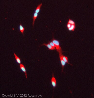 Immunocytochemistry/ Immunofluorescence - Anti-nkx6.3 antibody (ab93235)
ICC/IF image of ab93235 stained SK-N-SH cells. The cells were 100% methanol fixed (10 min) and then incubated in 1%BSA / 10% normal goat serum / 0.3M glycine in 0.1% PBS-Tween for 1h to permeabilise the cells and block non-specific protein-protein interactions. The cells were then incubated with the antibody (ab93235, 5µg/ml) overnight at +4°C. The secondary antibody (green) was ab96899, DyLight® 488 goat anti-rabbit IgG (H+L) used at a 1/250 dilution for 1h. Alexa Fluor® 594 WGA was used to label plasma membranes (red) at a 1/200 dilution for 1h. DAPI was used to stain the cell nuclei (blue) at a concentration of 1.43µM.
Protocols
Datasheets and documents
References (0)
ab93235 has not yet been referenced specifically in any publications.
Images
-
Western blot - Anti-nkx6.3 antibody (ab93235)Anti-nkx6.3 antibody (ab93235) at 1 µg/ml + E12 Mouse Embryo Brain and Spinal Cord Tissue Lysate at 10 µg
Secondary
Goat polyclonal Secondary Antibody to Rabbit IgG - H&L (HRP), pre-adsorbed at 1/5000 dilution
Developed using the ECL technique.
Performed under reducing conditions.
Predicted band size: 28 kDa
Observed band size: 28 kDa
Additional bands at: 38 kDa, 42 kDa, 90 kDa. We are unsure as to the identity of these extra bands.
Exposure time: 12 minutes
-
Immunocytochemistry/ Immunofluorescence - Anti-nkx6.3 antibody (ab93235)
ICC/IF image of ab93235 stained SK-N-SH cells. The cells were 100% methanol fixed (10 min) and then incubated in 1%BSA / 10% normal goat serum / 0.3M glycine in 0.1% PBS-Tween for 1h to permeabilise the cells and block non-specific protein-protein interactions. The cells were then incubated with the antibody (ab93235, 5µg/ml) overnight at +4°C. The secondary antibody (green) was ab96899, DyLight® 488 goat anti-rabbit IgG (H+L) used at a 1/250 dilution for 1h. Alexa Fluor® 594 WGA was used to label plasma membranes (red) at a 1/200 dilution for 1h. DAPI was used to stain the cell nuclei (blue) at a concentration of 1.43µM.


