Anti-Neurofilament heavy polypeptide antibody (ab4680)
")
Key features and details
- Chicken polyclonal to Neurofilament heavy polypeptide
- Suitable for: ICC, IHC-FrFl, WB
- Reacts with: Mouse, Rat, Cow, Human
- Isotype: IgY
Overview
-
Product name
Anti-Neurofilament heavy polypeptide antibody
See all Neurofilament heavy polypeptide primary antibodies -
Description
Chicken polyclonal to Neurofilament heavy polypeptide -
Host species
Chicken -
Tested Applications & Species
See all applications and species dataApplication Species ICC RatHumanIHC-FrFl RatWB Rat
-
Immunogen
Full length native protein (cow intermediate filaments were prepared from spinal cords by the glycerol polymerization method of Delacourte et al., and the cytoskeletal material was dissolved in 6M urea. Individual neurofilament subunits were purified by ion exchange chromatography on DEAE-cellulose and the NF-H containing fractions were concentrated and further purified by preparative gel electrophoresis on a Biorad Prepcell).
Properties
-
Form
Liquid -
Storage instructions
Shipped at 4°C. Store at +4°C. Do Not Freeze. -
Storage buffer
Preservative: 0.032% Sodium azide
Constituent: PBS -
 Concentration information loading...
 -
Purity
IgY fraction -
Purification notes
This is an IgY prep, similar to a mammalian ammonium sulphate IgG prep from serum. It is virtually pure IgY and we estimate that about 5% of material is specific to the immunogen. -
Clonality
Polyclonal -
Isotype
IgY -
Research areas
Images
-
Immunohistochemistry - Free Floating - Anti-Neurofilament heavy polypeptide antibody (ab4680)
Immunohistological analysis of a rat cerebellum section stained with ab4680 at a dilution 1:5,000 in red, and co-stained with rabbit pAb to GFAP at a dilution 1:5,000 in green. The blue is DAPI staining of nuclear DNA.
Following transcardial perfusion with 4% paraformaldehyde, brain was post fixed for 24 hours, cut to 45 μM, and free floating sections were stained.
-
 Western blot - Anti-Neurofilament heavy polypeptide antibody (ab4680)All lanes : Anti-Neurofilament heavy polypeptide antibody (ab4680) at 1/20000 dilution
Lane 1 : Rat spinal cord lysate
Lane 2 : Mouse spinal cord lysate
Lane 3 : Cow spinal cord lysate
Predicted band size: 200-220 kDa
-
 Immunocytochemistry - Anti-Neurofilament heavy polypeptide antibody (ab4680)
Mixed neuron/glial cultures stained with a mouse monoclonal antibody to neurofilament subunit NF-L (green) and ab4680, chicken antibody to neurofilament heavy polypeptide. This antibody binds primarily to the phosphorylated axonal forms of NF-H, in contrast to the NF-L antibody which stains both axonal and dendritic/perikaryal neurofilaments. The NF-L antibody therefore reveals a prominent cell body in green, while the surrounding axonal profiles are orange, since the are bound by both NF-L and the chicken NF-H antibody. Blue is a DNA stain.
-
 Immunocytochemistry - Anti-Neurofilament heavy polypeptide antibody (ab4680)Human neuroblastoma SH-SY5Y cells stained with chicken anti-NF-H (red) and mouse monoclonal to fibrillarin 38F3 (green). Nuclear DNA is revealed with Hoechst dye (blue). The NF-H antibody was used at a dilution of 1/100000 and the fibrillarin monoclonal at 1/1000. Cultures were processed using our standard fixation and staining procedure (in protocol section).
 -
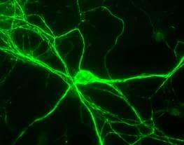 Immunocytochemistry - Anti-Neurofilament heavy polypeptide antibody (ab4680)
Rat Neurons stained with chicken anti-NF-H (green). The NF-H antibody was used at a dilution of 1/100000 using our standard fixation and staining procedure (in protocol section).
-
 Immunocytochemistry - Anti-Neurofilament heavy polypeptide antibody (ab4680) Image from Wirt SE et al, J Cell Biol. 2010 Nov 15;191(4):809-25. Epub 2010 Nov 8, Fig 4. DOI 10.1083/jcb.201003048
ab4680 staining Neurofilament heavy polypeptide (green) in murine embryoid bodies by Immunocytochemistry/ Immunofluorescence. Nuclei were visualized with DAPI.


