Anti-Mineralocorticoid Receptor antibody (ab64457)
Key features and details
- Rabbit polyclonal to Mineralocorticoid Receptor
- Suitable for: WB, IHC-P, ICC/IF
- Reacts with: Mouse, Human
- Isotype: IgG
Overview
-
Product name
Anti-Mineralocorticoid Receptor antibody
See all Mineralocorticoid Receptor primary antibodies -
Description
Rabbit polyclonal to Mineralocorticoid Receptor -
Host species
Rabbit -
Tested applications
Suitable for: WB, IHC-P, ICC/IFmore details -
Species reactivity
Reacts with: Mouse, Human
Predicted to work with: Rat, Xenopus laevis, Non human primates
-
Immunogen
Synthetic peptide conjugated to KLH derived from within residues 950 to the C-terminus of Human Mineralocorticoid Receptor.
Read Abcam's proprietary immunogen policy (Peptide available as ab74464.) -
Positive control
- This antibody gave a positive signal in the following lysates: Mouse Kidney Tissue Human Small Intestine Tissue Human Colon Tissue
-
General notes
Reproducibility is key to advancing scientific discovery and accelerating scientists’ next breakthrough.
Abcam is leading the way with our range of recombinant antibodies, knockout-validated antibodies and knockout cell lines, all of which support improved reproducibility.
We are also planning to innovate the way in which we present recommended applications and species on our product datasheets, so that only applications & species that have been tested in our own labs, our suppliers or by selected trusted collaborators are covered by our Abpromise™ guarantee.
In preparation for this, we have started to update the applications & species that this product is Abpromise guaranteed for.
We are also updating the applications & species that this product has been “predicted to work with,” however this information is not covered by our Abpromise guarantee.
Applications & species from publications and Abreviews that have not been tested in our own labs or in those of our suppliers are not covered by the Abpromise guarantee.
Please check that this product meets your needs before purchasing. If you have any questions, special requirements or concerns, please send us an inquiry and/or contact our Support team ahead of purchase. Recommended alternatives for this product can be found below, as well as customer reviews and Q&As.
Properties
-
Form
Liquid -
Storage instructions
Shipped at 4°C. Store at +4°C short term (1-2 weeks). Upon delivery aliquot. Store at -20°C or -80°C. Avoid freeze / thaw cycle. -
Storage buffer
pH: 7.40
Preservative: 0.02% Sodium azide
Constituent: PBS
Batches of this product that have a concentration Concentration information loading...
Purity
Immunogen affinity purifiedClonality
PolyclonalIsotype
IgGResearch areas
Associated products
-
Compatible Secondaries
-
Immunizing Peptide (Blocking)
-
Isotype control
Applications
Our Abpromise guarantee covers the use of ab64457 in the following tested applications.
The application notes include recommended starting dilutions; optimal dilutions/concentrations should be determined by the end user.
Application Abreviews Notes WB Use a concentration of 1 µg/ml. Detects a band of approximately 100 kDa (predicted molecular weight: 107 kDa). IHC-P Use a concentration of 1 µg/ml. ICC/IF Use a concentration of 1 µg/ml. Target
-
Function
Receptor for both mineralocorticoids (MC) such as aldosterone and glucocorticoids (GC) such as corticosterone or cortisol. Binds to mineralocorticoid response elements (MRE) and transactivates target genes. The effect of MC is to increase ion and water transport and thus raise extracellular fluid volume and blood pressure and lower potassium levels. -
Tissue specificity
Ubiquitous. Highly expressed in distal tubules, convoluted tubules and cortical collecting duct in kidney, and in sweat glands. Detected at lower levels in cardiomyocytes, in epidermis and in colon enterocytes. -
Involvement in disease
Defects in NR3C2 are a cause of autosomal dominant pseudohypoaldosteronism type I (AD-PHA1) [MIM:177735]. PHA1 is characterized by urinary salt wasting, resulting from target organ unresponsiveness to mineralocorticoids. There are 2 forms of PHA1: the autosomal dominant form that is mild, and the recessive form which is more severe and due to defects in any of the epithelial sodium channel subunits. In AD-PHA1 the target organ defect is confined to kidney. Clinical expression can vary from asymptomatic to moderate. It may be severe at birth, but symptoms remit with age. Familial and sporadic cases have been reported.
Defects in NR3C2 are a cause of early-onset hypertension with severe exacerbation in pregnancy (EOHSEP) [MIM:605115]. Inheritance is autosomal dominant. The disease is characterized by the onset of severe hypertension before the age of 20, and by suppression of aldosterone secretion. -
Sequence similarities
Belongs to the nuclear hormone receptor family. NR3 subfamily.
Contains 1 nuclear receptor DNA-binding domain. -
Domain
Composed of three domains: a modulating N-terminal domain, a DNA-binding domain and a C-terminal ligand-binding domain. -
Post-translational
modificationsPhosphorylated. -
Cellular localization
Cytoplasm. Nucleus. Endoplasmic reticulum membrane. Cytoplasmic and nuclear in the absence of ligand; nuclear after ligand-binding. When bound to HSD11B2, it is found associated with the endoplasmic reticulum membrane. - Information by UniProt
-
Database links
- Entrez Gene: 4306 Human
- Entrez Gene: 110784 Mouse
- Entrez Gene: 25672 Rat
- Entrez Gene: 399290 Xenopus laevis
- Omim: 600983 Human
- SwissProt: P08235 Human
- SwissProt: Q8VII8 Mouse
- SwissProt: P22199 Rat
see all -
Alternative names
- Aldosterone receptor antibody
- MCR antibody
- MCR_HUMAN antibody
see all
Images
-
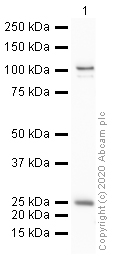
Anti-Mineralocorticoid Receptor antibody (ab64457) at 1 µg/ml + Human small intestine tissue lysate - total protein at 10 µg
Secondary
Goat polyclonal to Rabbit IgG - H&L - Pre-Adsorbed (HRP) at 1/3000 dilution
Developed using the ECL technique.
Performed under reducing conditions.
Predicted band size: 107 kDa
Observed band size: 102 kDa why is the actual band size different from the predicted?
Exposure time: 1 minuteThis blot was produced using a 4-12% Bis-tris gel under the MOPS buffer system. The gel was run at 200V for 50 minutes before being transferred onto a Nitrocellulose membrane at 30V for 70 minutes. The membrane was then blocked for an hour using 2% Bovine Serum Albumin before being incubated with ab64457 overnight at 4°C. Antibody binding was detected using an anti-rabbit antibody conjugated to HRP, and visualised using ECL development solution ab133406.
-
Anti-Mineralocorticoid Receptor antibody (ab64457) at 1 µg/ml + Kidney (Mouse) Tissue Lysate at 10 µg
Secondary
Goat polyclonal to Rabbit IgG - H&L - Pre-Adsorbed (HRP) at 1/3000 dilution
Developed using the ECL technique.
Performed under reducing conditions.
Predicted band size: 107 kDa
Observed band size: 102 kDa why is the actual band size different from the predicted?
Additional bands at: 200 kDa (possible non-specific binding), 87 kDa (possible non-specific binding), 95 kDa (possible non-specific binding)
Exposure time: 12 minutesThis blot was produced using a 4-12% Bis-tris gel under the MOPS buffer system. The gel was run at 200V for 50 minutes before being transferred onto a Nitrocellulose membrane at 30V for 70 minutes. The membrane was then blocked for an hour using 2% Bovine Serum Albumin before being incubated with ab64457 overnight at 4°C. Antibody binding was detected using an anti-rabbit antibody conjugated to HRP, and visualised using ECL development solution ab133406.
-
ICC/IF image of ab64457 stained HeLa cells. The cells were 4% PFA fixed (10 min) and then incubated in 1%BSA / 10% normal goat serum / 0.3M glycine in 0.1% PBS-Tween for 1h to permeabilise the cells and block non-specific protein-protein interactions. The cells were then incubated with the antibody (ab64457, 1µg/ml) overnight at +4°C. The secondary antibody (green) was Alexa Fluor® 488 goat anti-rabbit IgG (H+L) used at a 1/1000 dilution for 1h. Alexa Fluor® 594 WGA was used to label plasma membranes (red) at a 1/200 dilution for 1h. DAPI was used to stain the cell nuclei (blue) at a concentration of 1.43µM. This antibody also gave a positive result in 4% PFA fixed (10 min) HepG2, Hek293 and MCF7 cells at 1µg/ml, and in 100% methanol fixed (5 min) HeLa, Hek293, HepG2 and MCF7 cells at 1µg/ml.
-
 Immunohistochemistry (Formalin/PFA-fixed paraffin-embedded sections) - Anti-Mineralocorticoid Receptor antibody (ab64457)IHC image of Mineralocorticoid Receptor staining in Human Tonsil FFPE section, performed on a BondTM system using the standard protocol F. The section was pre-treated using heat mediated antigen retrieval with sodium citrate buffer (pH6, epitope retrieval solution 1) for 20 mins. The section was then incubated with ab64457, 1µg/ml, for 15 mins at room temperature and detected using an HRP conjugated compact polymer system. DAB was used as the chromogen. The section was then counterstained with haematoxylin and mounted with DPX
Protocols
Datasheets and documents
References (12)
ab64457 has been referenced in 12 publications.
- Song X et al. Zi Shen Huo Luo Formula Enhances the Therapeutic Effects of Angiotensin-Converting Enzyme Inhibitors on Hypertensive Left Ventricular Hypertrophy by Interfering With Aldosterone Breakthrough and Affecting Caveolin-1/Mineralocorticoid Receptor Colocalization and Downstream Extracellular Signal-Regulated Kinase Signaling. Front Pharmacol 11:383 (2020). PubMed: 32317965
- Chen J et al. Paeoniflorin regulates the hypothalamic-pituitary-adrenal axis negative feedback in a rat model of post-traumatic stress disorder. Iran J Basic Med Sci 23:439-448 (2020). PubMed: 32489558
- Yang C et al. MicroRNA-766 promotes cancer progression by targeting NR3C2 in hepatocellular carcinoma. FASEB J 33:1456-1467 (2019). PubMed: 30130435
- Wang W et al. Ketamine improved depressive-like behaviors via hippocampal glucocorticoid receptor in chronic stress induced- susceptible mice. Behav Brain Res 364:75-84 (2019). PubMed: 30753876
- Hayakawa K et al. MicroRNA-766-3p Contributes to Anti-Inflammatory Responses through the Indirect Inhibition of NF-?B Signaling. Int J Mol Sci 20:N/A (2019). PubMed: 30769772
Images
-
Anti-Mineralocorticoid Receptor antibody (ab64457) at 1 µg/ml + Human small intestine tissue lysate - total protein at 10 µg
Secondary
Goat polyclonal to Rabbit IgG - H&L - Pre-Adsorbed (HRP) at 1/3000 dilution
Developed using the ECL technique.
Performed under reducing conditions.
Predicted band size: 107 kDa
Observed band size: 102 kDa why is the actual band size different from the predicted?
Exposure time: 1 minuteThis blot was produced using a 4-12% Bis-tris gel under the MOPS buffer system. The gel was run at 200V for 50 minutes before being transferred onto a Nitrocellulose membrane at 30V for 70 minutes. The membrane was then blocked for an hour using 2% Bovine Serum Albumin before being incubated with ab64457 overnight at 4°C. Antibody binding was detected using an anti-rabbit antibody conjugated to HRP, and visualised using ECL development solution ab133406.
-
Anti-Mineralocorticoid Receptor antibody (ab64457) at 1 µg/ml + Kidney (Mouse) Tissue Lysate at 10 µg
Secondary
Goat polyclonal to Rabbit IgG - H&L - Pre-Adsorbed (HRP) at 1/3000 dilution
Developed using the ECL technique.
Performed under reducing conditions.
Predicted band size: 107 kDa
Observed band size: 102 kDa why is the actual band size different from the predicted?
Additional bands at: 200 kDa (possible non-specific binding), 87 kDa (possible non-specific binding), 95 kDa (possible non-specific binding)
Exposure time: 12 minutesThis blot was produced using a 4-12% Bis-tris gel under the MOPS buffer system. The gel was run at 200V for 50 minutes before being transferred onto a Nitrocellulose membrane at 30V for 70 minutes. The membrane was then blocked for an hour using 2% Bovine Serum Albumin before being incubated with ab64457 overnight at 4°C. Antibody binding was detected using an anti-rabbit antibody conjugated to HRP, and visualised using ECL development solution ab133406.
-
ICC/IF image of ab64457 stained HeLa cells. The cells were 4% PFA fixed (10 min) and then incubated in 1%BSA / 10% normal goat serum / 0.3M glycine in 0.1% PBS-Tween for 1h to permeabilise the cells and block non-specific protein-protein interactions. The cells were then incubated with the antibody (ab64457, 1µg/ml) overnight at +4°C. The secondary antibody (green) was Alexa Fluor® 488 goat anti-rabbit IgG (H+L) used at a 1/1000 dilution for 1h. Alexa Fluor® 594 WGA was used to label plasma membranes (red) at a 1/200 dilution for 1h. DAPI was used to stain the cell nuclei (blue) at a concentration of 1.43µM. This antibody also gave a positive result in 4% PFA fixed (10 min) HepG2, Hek293 and MCF7 cells at 1µg/ml, and in 100% methanol fixed (5 min) HeLa, Hek293, HepG2 and MCF7 cells at 1µg/ml.
-
 Immunohistochemistry (Formalin/PFA-fixed paraffin-embedded sections) - Anti-Mineralocorticoid Receptor antibody (ab64457)IHC image of Mineralocorticoid Receptor staining in Human Tonsil FFPE section, performed on a BondTM system using the standard protocol F. The section was pre-treated using heat mediated antigen retrieval with sodium citrate buffer (pH6, epitope retrieval solution 1) for 20 mins. The section was then incubated with ab64457, 1µg/ml, for 15 mins at room temperature and detected using an HRP conjugated compact polymer system. DAB was used as the chromogen. The section was then counterstained with haematoxylin and mounted with DPX


