Anti-LATS1/WARTS antibody (ab234820)
")
Key features and details
- Rabbit polyclonal to LATS1/WARTS
- Suitable for: IHC-P, ICC/IF, IP
- Reacts with: Human
- Isotype: IgG
Overview
-
Product name
Anti-LATS1/WARTS antibody
See all LATS1/WARTS primary antibodies -
Description
Rabbit polyclonal to LATS1/WARTS -
Host species
Rabbit -
Tested applications
Suitable for: IHC-P, ICC/IF, IPmore details -
Species reactivity
Reacts with: Human -
Immunogen
Recombinant fragment corresponding to Human LATS1/WARTS aa 1-280.
Database link: O95835 -
Positive control
- IHC-P: Human liver cancer, placenta and adrenal gland tissues. ICC/IF: HeLa cells. IP: K562 cell lysate.
-
General notes
This product was previously labelled as LATS1
Reproducibility is key to advancing scientific discovery and accelerating scientists’ next breakthrough.
Abcam is leading the way with our range of recombinant antibodies, knockout-validated antibodies and knockout cell lines, all of which support improved reproducibility.
We are also planning to innovate the way in which we present recommended applications and species on our product datasheets, so that only applications & species that have been tested in our own labs, our suppliers or by selected trusted collaborators are covered by our Abpromise™ guarantee.
In preparation for this, we have started to update the applications & species that this product is Abpromise guaranteed for.
We are also updating the applications & species that this product has been “predicted to work with,” however this information is not covered by our Abpromise guarantee.
Applications & species from publications and Abreviews that have not been tested in our own labs or in those of our suppliers are not covered by the Abpromise guarantee.
Please check that this product meets your needs before purchasing. If you have any questions, special requirements or concerns, please send us an inquiry and/or contact our Support team ahead of purchase. Recommended alternatives for this product can be found below, as well as customer reviews and Q&As.
Properties
-
Form
Liquid -
Storage instructions
Shipped at 4°C. Store at +4°C short term (1-2 weeks). Upon delivery aliquot. Store at -20°C long term. Avoid freeze / thaw cycle. -
Storage buffer
pH: 7.40
Constituents: 50% Glycerol (glycerin, glycerine), PBS, 0.03% Proclin 300 -
 Concentration information loading...
 -
Purity
Protein G purified -
Purification notes
Purity >95%. -
Clonality
Polyclonal -
Isotype
IgG -
Research areas
Images
-
Immunocytochemistry/ Immunofluorescence - Anti-LATS1/WARTS antibody (ab234820)
HeLa (human epithelial cell line from cervix adenocarcinoma) cells stained for LATS1/WARTS (green) using ab234820 at 1/100 dilution in ICC/IF, followed by Alexa Fluor 488® congugated Goat Anti-Rabbit IgG (H+L).
-
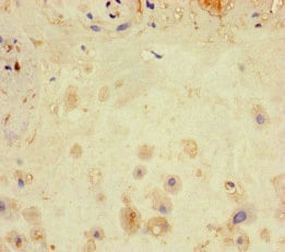
 Immunohistochemistry (Formalin/PFA-fixed paraffin-embedded sections) - Anti-LATS1/WARTS antibody (ab234820)
Paraffin-embedded human liver cancer tissue stained for LATS1/WARTS using ab234820 at 1/100 dilution in immunohistochemical analysis.
-
 Immunoprecipitation - Anti-LATS1/WARTS antibody (ab234820)
LATS1/WARTS was immunoprecipitated from 500 µg K562 (human chronic myelogenous leukemia cell line from bone marrow) whole cell lysate with 6 µg ab234820.
Lane 1: Rabbit control IgG IP (1 µg) in K562 whole cell lysate.
Lane 2: ab234820 IP in K562 whole cell lysate.
Lane 3: K562 whole cell lysate 10 µg (Input).For western blotting, a HRP-conjugated Protein G antibody was used as the secondary antibody at 1/2000 dilution.
-
 Immunohistochemistry (Formalin/PFA-fixed paraffin-embedded sections) - Anti-LATS1/WARTS antibody (ab234820)
Paraffin-embedded human placenta tissue stained for LATS1/WARTS using ab234820 at 1/100 dilution in immunohistochemical analysis.
-
 Immunohistochemistry (Formalin/PFA-fixed paraffin-embedded sections) - Anti-LATS1/WARTS antibody (ab234820)
Paraffin-embedded human liver cancer tissue stained for LATS1/WARTS using ab234820 at 1/100 dilution in immunohistochemical analysis.
Performed on a Leica BondTM system. After dewaxing and hydration, antigen retrieval was mediated by high pressure in a citrate buffer (pH 6.0). Section was blocked with 10% normal goat serum 30min at RT. Then primary antibody (1% BSA) was incubated at 4°C overnight. The primary is detected by a biotinylated secondary antibody.
-
 Immunohistochemistry (Formalin/PFA-fixed paraffin-embedded sections) - Anti-LATS1/WARTS antibody (ab234820)
Paraffin-embedded human adrenal gland tissue stained for LATS1/WARTS using ab234820 at 1/100 dilution in immunohistochemical analysis.
Performed on a Leica BondTM system. After dewaxing and hydration, antigen retrieval was mediated by high pressure in a citrate buffer (pH 6.0). Section was blocked with 10% normal goat serum 30min at RT. Then primary antibody (1% BSA) was incubated at 4°C overnight. The primary is detected by a biotinylated secondary antibody.


