Anti-KMT2A / MLL antibody (ab272023)
Key features and details
- Rabbit polyclonal to KMT2A / MLL
- Suitable for: WB, IP, ChIP/Chip
- Reacts with: Human
- Isotype: IgG
Overview
-
Product name
Anti-KMT2A / MLL antibody
See all KMT2A / MLL primary antibodies -
Description
Rabbit polyclonal to KMT2A / MLL -
Host species
Rabbit -
Tested applications
Suitable for: WB, IP, ChIP/Chipmore details -
Species reactivity
Reacts with: Human
Predicted to work with: Mouse
-
Immunogen
Synthetic peptide within Human KMT2A/ MLL aa 2725-2775. The exact sequence is proprietary. The epitope is found in the C-terminal 180 kDa fragment generated by proteolytic cleavage. The epitope is found in isoform 14P-18B of KMT2A/ MLL.
Database link: Q03164 -
Positive control
- WB: HeLa, Jurkat and HEK-293T whole cell lysate. IP: HEK-293T whole cell lysate. ChIP on ChIP: Chromatin from K562 cells.
Properties
-
Form
Liquid -
Storage instructions
Shipped at 4°C. Store at +4°C short term (1-2 weeks). Upon delivery aliquot. Store at -20°C long term. Avoid freeze / thaw cycle. -
Storage buffer
pH: 7
Preservative: 0.09% Sodium azide
Constituent: Tris citrate/phosphate
pH 7 to 8 -
 Concentration information loading...
 -
Purity
Immunogen affinity purified -
Clonality
Polyclonal -
Isotype
IgG -
Research areas
Images
-
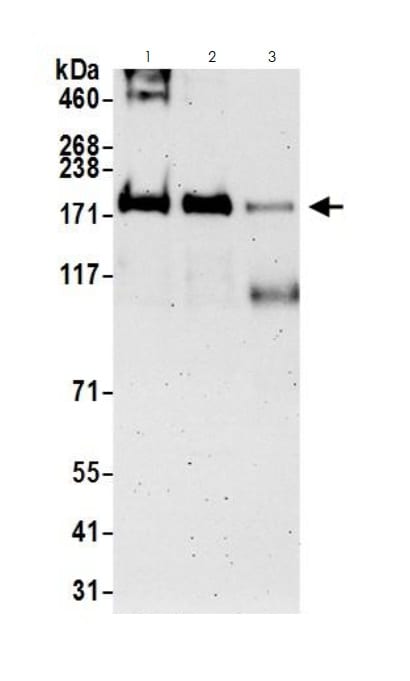
All lanes : Anti-KMT2A / MLL antibody (ab272023) at 0.1 µg/ml
Lane 1 : HeLa (Human epithelial cell line from cervix adenocarcinoma) whole cell lysate
Lane 2 : HEK-293T (Human epithelial cell line from embryonic kidney transformed with large T antigen) whole cell lysate
Lane 3 : Jurkat (Human T cell leukemia cell line from peripheral blood) whole cell lysate
Lysates/proteins at 50 µg per lane.
Predicted band size: 432 kDa
Exposure time: 3 minutes
-
KMT2A / MLL was immunoprecipitated from HEK-293T (Human epithelial cell line from embryonic kidney transformed with large T antigen) whole cell lysate (1 mg for IP, 20% of IP loaded) with ab272023 at 3 μg per reaction. WB was performed using ab272023 at 1 µg/ml.
Lane 1: ab272023 IP in HEK-293T whole cell lysate.
Lane 2: Control IgG IP.
Exposure time: 3 minutes. -
ChIP-chip scatter plot of ab272023 enriched DNA binding sites versus input reference DNA.
Plot A. 10 μg of ab272023 was used to immunoprecipitate chromatin from K562 cells according to Ren et al (Genes Dev. 2002 16: 245-256). Immunoprecipitatesd DNA and reference DNA were amplified via ligation-mediated PCR and the products labeled with fluorescent dUTPs. The labeled ChIP and reference DNA were pooled, hybridized to a DNA microarray, and analyzed. Data points below the +3 SD curve (red line) represent significantly enriched binding sites.
Plot B. As a control, a similar experiment was performed using normal rabbit IgG. Compared to the ab272023 ChIP, normal rabbit IgG showed little enrichment.


