Anti-Integrin linked ILK antibody (ab74336)
")
Key features and details
- Rabbit polyclonal to Integrin linked ILK
- Suitable for: WB, IP, ICC/IF
- Reacts with: Mouse, Human
- Isotype: IgG
Overview
-
Product name
Anti-Integrin linked ILK antibody
See all Integrin linked ILK primary antibodies -
Description
Rabbit polyclonal to Integrin linked ILK -
Host species
Rabbit -
Tested applications
Suitable for: WB, IP, ICC/IFmore details -
Species reactivity
Reacts with: Mouse, Human
Predicted to work with: Rat, Sheep, Rabbit, Horse, Chicken, Guinea pig, Cow, Dog, Pig, Chimpanzee, Zebrafish, Rhesus monkey, Gorilla, Tilapia, Orangutan, Medaka fish
-
Immunogen
Synthetic peptide corresponding to a region between residues 402 and 451 of human Integrin linked ILK (NP_004508.1)
-
Positive control
- HeLa, 293T and NIH3T3 whole cell lysates.
-
General notes
Reproducibility is key to advancing scientific discovery and accelerating scientists’ next breakthrough.
Abcam is leading the way with our range of recombinant antibodies, knockout-validated antibodies and knockout cell lines, all of which support improved reproducibility.
We are also planning to innovate the way in which we present recommended applications and species on our product datasheets, so that only applications & species that have been tested in our own labs, our suppliers or by selected trusted collaborators are covered by our Abpromise™ guarantee.
In preparation for this, we have started to update the applications & species that this product is Abpromise guaranteed for.
We are also updating the applications & species that this product has been “predicted to work with,” however this information is not covered by our Abpromise guarantee.
Applications & species from publications and Abreviews that have not been tested in our own labs or in those of our suppliers are not covered by the Abpromise guarantee.
Please check that this product meets your needs before purchasing. If you have any questions, special requirements or concerns, please send us an inquiry and/or contact our Support team ahead of purchase. Recommended alternatives for this product can be found below, as well as customer reviews and Q&As.
Properties
-
Form
Liquid -
Storage instructions
Shipped at 4°C. Upon delivery aliquot and store at -20°C. Avoid freeze / thaw cycles. -
Storage buffer
pH: 6.8
Preservative: 0.09% Sodium azide
Constituents: 0.1% BSA, Tris buffered saline -
 Concentration information loading...
 -
Purity
Immunogen affinity purified -
Purification notes
ab74336 was affinity purified using an epitope specific to Integrin linked ILK immobilized on solid support. -
Clonality
Polyclonal -
Isotype
IgG -
Research areas
Images
-
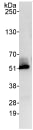
Western blot - Anti-Integrin linked ILK antibody (ab74336)All lanes : Anti-Integrin linked ILK antibody (ab74336) at 0.04 µg/ml
Lane 1 : HeLa whole cell lysate at 50 µg
Lane 2 : HeLa whole cell lysate at 15 µg
Lane 3 : HeLa whole cell lysate at 5 µg
Lane 4 : 293T whole cell lysate at 50 µg
Lane 5 : NIH3T3 whole cell lysate at 50 µg
Developed using the ECL technique.
Predicted band size: 51 kDa
Observed band size: 51 kDa
Exposure time: 10 seconds
-
 Immunocytochemistry/ Immunofluorescence - Anti-Integrin linked ILK antibody (ab74336)Immunocytochemistry/Immunofluorescence analysis of acetone-fixed asynchronous HeLa cells labelling Integrin linked ILK with ab74336 at 1/100 (2µg/ml). A red-fluorescent goat anti-rabbit IgG (1/100) was used as the secondary antibody.
 -
 Immunoprecipitation - Anti-Integrin linked ILK antibody (ab74336)Detection of Integrin linked ILK by Western Blot of Immunprecipitate.
ab74336 at 1µg/ml staining Integrin linked ILK in HeLa whole cell lysate immunoprecipitated using ab74336 at 3µg/mg lysate (1 mg/IP; 20% of IP loaded/lane).
Detection: Chemiluminescence with exposure time of 10 seconds. -
 Immunocytochemistry/ Immunofluorescence - Anti-Integrin linked ILK antibody (ab74336)ICC/IF image of ab74336 stained MCF7 cells. The cells were 100% methanol fixed (5 min) and then incubated in 1%BSA / 10% normal goat serum / 0.3M glycine in 0.1% PBS-Tween for 1h to permeabilise the cells and block non-specific protein-protein interactions. The cells were then incubated with the antibody (ab74336, 1µg/ml) overnight at +4°C. The secondary antibody (green) was Alexa Fluor® 488 goat anti-rabbit IgG (H+L) used at a 1/1000 dilution for 1h. Alexa Fluor® 594 WGA was used to label plasma membranes (red) at a 1/200 dilution for 1h. DAPI was used to stain the cell nuclei (blue) at a concentration of 1.43µM.


