Anti-IL-6 antibody (ab6672)
")
Key features and details
- Rabbit polyclonal to IL-6
- Suitable for: WB, IHC-P
- Reacts with: Mouse, Human
- Isotype: IgG
Overview
-
Product name
Anti-IL-6 antibody
See all IL-6 primary antibodies -
Description
Rabbit polyclonal to IL-6 -
Host species
Rabbit -
Specificity
ab6672 detects a band at 25 kDa in human lung tissue lysate and mouse spleen tissue lysate, however the signal in mouse tissue is significantly lower. It also binds strongly to a protein at ~55 kDa in human lung tissue extracts, which we believe represents a glycosylated form of IL6. ab6672 also detects several bands in human lung tissue lysate within the region of 30-40 kDa. These may represent heteromers of IL6. Please be aware that this product has low homology with the mouse and rat sequence of IL6 (Rat, 40%; Mouse 41%, UniProt blast) and we therefore cannot guarantee reactivity in these species. -
Tested Applications & Species
See all applications and species dataApplication Species IHC-P HumanWB MouseHuman
-
Immunogen
Recombinant full length protein corresponding to Human IL-6. Produced in E.coli.
Database link: P05231 -
Positive control
- Purchase matching WB positive control:Recombinant Human IL-6 protein
- WB: Recombinant Human IL6 protein (ab101044), lysate of 2 x 10
-
General notes
IL-6 synonyms: plasmacytoma growth factor (PCT-GF),interferon-a-2 (IFN-a2), monocyte derived human B cellgrowth factor, B cell stimulating factor (BSF-2),hepatocyte stimulating factor (HSF), and interleukinhybridoma/plasmacytoma-1 (IL-HP1).
Properties
-
Form
Liquid -
Storage instructions
Shipped at 4°C. Store at +4°C short term (1-2 weeks). Upon delivery aliquot. Store at -20°C or -80°C. Avoid freeze / thaw cycle. -
Storage buffer
Constituents: 0.42% Potassium phosphate, 0.87% Sodium chloride -
 Concentration information loading...
 -
Purity
Whole antiserum -
Clonality
Polyclonal -
Isotype
IgG -
Research areas
Images
-
Immunohistochemistry (Formalin/PFA-fixed paraffin-embedded sections) - Anti-IL-6 antibody (ab6672)
IHC image of IL6 staining in human lung formalin fixed paraffin embedded tissue section*, performed on a Leica Bond™ system using the standard protocol F. The section was pre-treated using heat mediated antigen retrieval with sodium citrate buffer (pH6, epitope retrieval solution 1) for 20 mins. The section was then incubated with ab6672, 1/400, for 15 mins at room temperature and detected using an HRP conjugated compact polymer system. DAB was used as the chromogen. The section was then counterstained with haematoxylin and mounted with DPX.
For other IHC staining systems (automated and non-automated) customers should optimize variable parameters such as antigen retrieval conditions, primary antibody concentration and antibody incubation times.
*Tissue obtained from the Human Research Tissue Bank, supported by the NIHR Cambridge Biomedical Research Centre
-
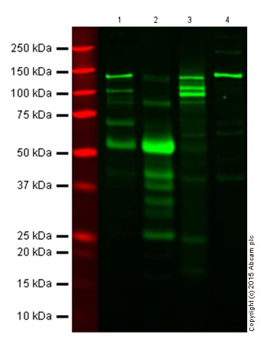 Western blot - Anti-IL-6 antibody (ab6672)All lanes : Anti-IL-6 antibody (ab6672) at 1/500 dilution
Lane 1 : Human spleen tissue lysate
Lane 2 : Human lung tissue lysate
Lane 3 : Mouse spleen tissue lysate
Lane 4 : Mouse lung tissue lysate
Lysates/proteins at 20 µg per lane.
Performed under reducing conditions.
Additional bands at: 25 kDa, 55 kDa (possible glycosylated form). We are unsure as to the identity of these extra bands.Tissue lysates were denatured for 10-15 minutes at 90ºC. ab6672 was incubated overnight at 4ºC and the secondary antibody for 1 hour at RT.
ab6672 detects a band at 25 kDa in Human lung tissue lysate and Mouse spleen tissue lysate, however the signal in mouse tissue is significantly lower. It also binds strongly to a protein at ~55 kDa in Human lung tissue extracts, which we believe represents a glycosylated form of IL6. ab6672 also detects several bands in Human lung tissue lysate within the region of 30-40 kDa. These may represent heteromers of IL6.
-
 Immunohistochemistry (Formalin/PFA-fixed paraffin-embedded sections) - Anti-IL-6 antibody (ab6672) Lee EJ et al. Radiation Inhibits Interleukin-12 Production via Inhibition of C-Rel through the Interleukin-6/ Signal Transducer and Activator of Transcription 3 Signaling Pathway in Dendritic Cells. PLoS One 11:e0146463 (2016).
IL6 (Red) and IL-12 (Green) were measured at 1, 3, and 7 days after 10 Gy irradiation of HCa-1 tumors to determine whether irradiation regulates IL-12 and IL6 expression in tumours. ab6672 was used to stain IL6 at 1/100 dilution in immunohistochemical analysis.
-
 Western blot - Anti-IL-6 antibody (ab6672)Anti-IL-6 antibody (ab6672) at 1/500 dilution + recombinant human IL-6
Secondary
conjugated anti-Rabbit IgG at 1/40000 dilution
Developed using the ECL technique.
Observed band size: 21 kDa why is the actual band size different from the predicted?4-20% Tris-Glycine gel.
The membrane was blocked for 30 minutes with 1% BSA-TBST.


