Anti-Histone H3 (tri methyl K56) antibody (ab272162)
Key features and details
- Rabbit polyclonal to Histone H3 (tri methyl K56)
- Suitable for: WB, Dot blot
- Reacts with: Human, Caenorhabditis elegans
- Isotype: IgG
Overview
-
Product name
Anti-Histone H3 (tri methyl K56) antibody
See all Histone H3 primary antibodies -
Description
Rabbit polyclonal to Histone H3 (tri methyl K56) -
Host species
Rabbit -
Tested Applications & Species
See all applications and species dataApplication Species Dot Recombinant fragmentIF HumanWB Caenorhabditis elegans -
Immunogen
Synthetic peptide corresponding to Human Histone H3 (tri methyl K56).
Database link: Q71DI3 -
Positive control
- WB: Caenorhabditis elegans embryo lysate. ICC: HeLa cells (prophase and telophase).
Properties
-
Form
Liquid -
Storage instructions
Shipped at 4°C. Store at +4°C short term (1-2 weeks). Upon delivery aliquot. Store at -20°C. Avoid freeze / thaw cycle. -
Storage buffer
Preservative: 0.01% Sodium azide
Constituents: 0.0004% Potassium phosphate, 0.0008% Sodium chloride, 30% Glycerol (glycerin, glycerine) -
 Concentration information loading...
 -
Purity
Immunogen affinity purified -
Clonality
Polyclonal -
Isotype
IgG -
Research areas
Images
-
Immunofluorescence analysis of ab272162. HeLa (Human epithelial cell line from cervix adenocarcinoma) cells are shown during prophase. Primary antibody ab272162 was used at 1:100 dilution for 1 hour at room temperature. Secondary antibody, Dylight 488 antibody at 1:10,000 for 45 minutes at room temperature. Histone H3 [Trimethyl Lys56] localization is nuclear and chromosomal and it is expressed in green, nuclei and alpha-tubulin are counterstained with DAPI (blue) and Dylight 550 (red). Fixation was 0.5% PFA. Antigen retrieval was not required.
-
Anti-Histone H3 (tri methyl K56) antibody (ab272162) at 1 µg/ml + Caenorhabditis elegans embryo lysate at 30 µg
Secondary
IRDye800™ rabbit secondary antibody at 1/10000 dilution
Predicted band size: 15 kDaBlock: 5% BLOTTO overnight at 4°C.
Predicted/Observed size: ~15 kDa.
Other band(s): None.
-
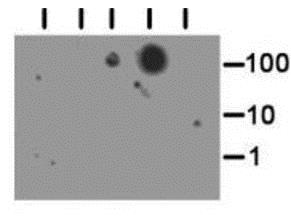
Dot Blot analysis of ab272162.
Lane 1: Ac.
Lane 2: me1.
Lane 3: me2.
Lane 4: me3.
Lane 5: unmodified.
Load: 1, 10, and 100 picomoles of peptide. Primary antibody ab272162 was used at 1:40 for 45 minutes at 4°C. Secondary antibody Dylight™488 rabbit antibody at 1:10,000 for 45 minutes at room temperature. Block: 5% BLOTTO overnight at 4°C.
-
Immunofluorescence of ab272162. HeLa (Human epithelial cell line from cervix adenocarcinoma) cells are shown during telophase. Primary antibody ab272162 was at a 1:100 dilution for 1 hour at room temperature. Dylight 488 secondary antibody followed at 1:10,000 for 45 minutes at toom temperature. Histone H3 [Trimethyl Lys56] localization is nuclear and chromosomal. Histone H3 [Trimethyl Lys56] staining is expressed in green, nuclei and alpha-tubulin are counterstained with DAPI (blue) and Dylight 550 (red). Fixation was 0.5% PFA and antigen retrieval was not required.


